)
)
.
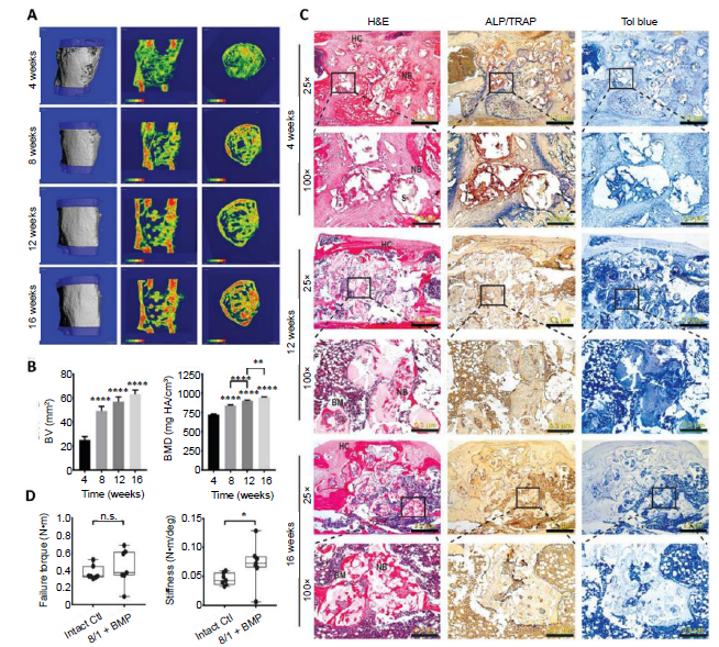
Figure 3.. Accelerated healing of 5-mm rat femoral segmental defects by 25% HAp-PELGA(8/1) grafts preabsorbed with 400-ng rhBMP-2/7. (A) 3D μCT images and BMD colour maps (centre sagittal and axial slices) of the ROI showing maturing regenerated bone within the defect over time. Global thresholding was applied to exclude bone densities below 518.2 mg HAp/cm3 (HAp-PELGA graft invisible at this threshold). (B) Longitudinal μCT quantification of BV and BMD (n ≥ 12) within the ROI over time. Data are presented as means ± SEM. **P < 0.01, ****P < 0.0001 (one-way analysis of variance with Tukey’s post-hoc test). The global lower threshold of 518.2 mg HAp/cm3 was applied for all quantifications. (C) Histological micrographs of H&E-, ALP/TRAP-, and Tol blue-stained sections of explanted graft-filled femurs over time. Scale bars: 1.2 mm (25× magnification) and 300 μm (100× magnification). Boxed regions shown at higher magnification in bottom rows. (D) Boxplots of failure torque and stiffness of intact (control) versus regenerated femur (8/1 + BMP) 16 weeks after being treated with HAp-PELGA(8/1) grafts preloaded with 400-ng rhBMP-2/7 (n = 7). *P < 0.05 (Wilcoxon-Mann-Whitney rank sum test). Reprinted from Zhang et al.