Three-dimensional biofabrication of an aragonite-enriched self-hardening bone graft substitute and assessment of its osteogenicity in vitro and in vivo
Yunsong Shi1,2, Ruijun He1, Xiangyu Deng1, Zengwu Shao1, Davide Deganello3, Chunze Yan4,*( ), Zhidao Xia2,*()
), Zhidao Xia2,*()
.
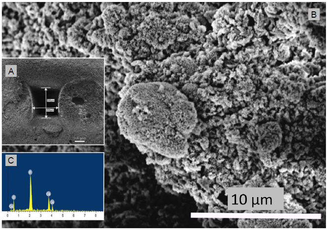
Figure 3.. (A) Scanning electron microscopic image of the cross section of hydroxyapatite/aragonite and measurement of pore size. (B) Higher magnification of the scanning electron microscopic image shown in A. (C) Energy dispersive X-ray spectroscopy analysis of the cross section of hydroxyapatite/aragonite. Scale bars: 100 μm in A, 10 μm in B.