Design and evaluation of a novel sub-scaffold dental implant system based on the osteoinduction of micro-nano bioactive glass
Fujian Zhao1, Zhen Yang2,3, Lu Liu2,3, Dafu Chen4, Longquan Shao1,*( ), Xiaofeng Chen2,3,*()
), Xiaofeng Chen2,3,*()
.
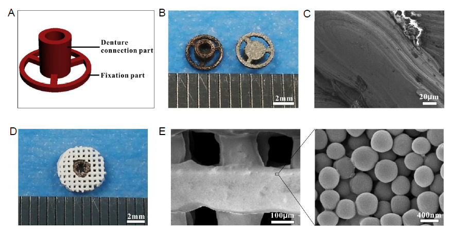
Figure 1.. Composition and characterization of the SDIS. (A) A CAD model of the metal implant which consists of two parts: a denture connection part and a fixation part. (B) Digital photos of the two opposite surfaces of the metal implant, created by the SLM process. (C) SEM image of the metal implant. (D) Digital photo of the SDIS created by assembling the metal implant and MNBG scaffold together. (E) SEM image of the MNBG scaffold. Scale bars: 2 mm in B and D, 20 μm in C, 200 μm in E, 400 nm in enlarge part. CAD: computer-aided design; MNBG: micro-nano bioactive glass; SDIS: scaffold dental implant system; SEM: scanning electron microscopy; SLM: selective laser melting.