Design and evaluation of a novel sub-scaffold dental implant system based on the osteoinduction of micro-nano bioactive glass
Fujian Zhao1, Zhen Yang2,3, Lu Liu2,3, Dafu Chen4, Longquan Shao1,*( ), Xiaofeng Chen2,3,*()
), Xiaofeng Chen2,3,*()
.
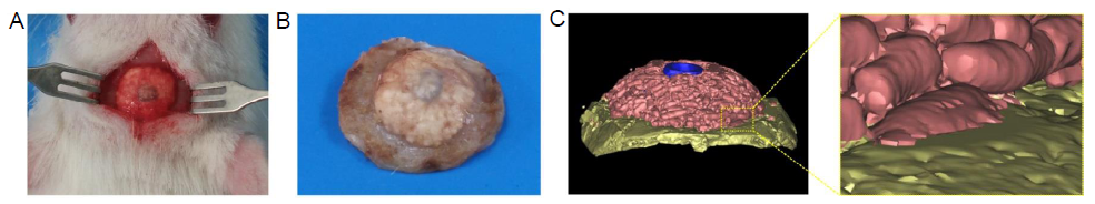
Figure 3.. (A) Digital photo of the reparative effect. (B) Residual SDIS and surrounding bone tissue at week 6. (C) Micro-CT analysis of 3-dimensional reconstructed images of SDIS and surrounding tissue after implantation for 6 weeks. A magnified image of the join between implant and bone, showed good integration. Pink indicates residual bioactive glass scaffold, blue indicates the metal implant, and yellow indicates the skull. SDIS: scaffold dental implant system.