Biofunctional magnesium coating of implant materials by physical vapour deposition
Qingchuan Wang1, Weidan Wang1,2, Yanfang Li3, Weirong Li3, Lili Tan1,*( ), Ke Yang1,*()
), Ke Yang1,*()
.
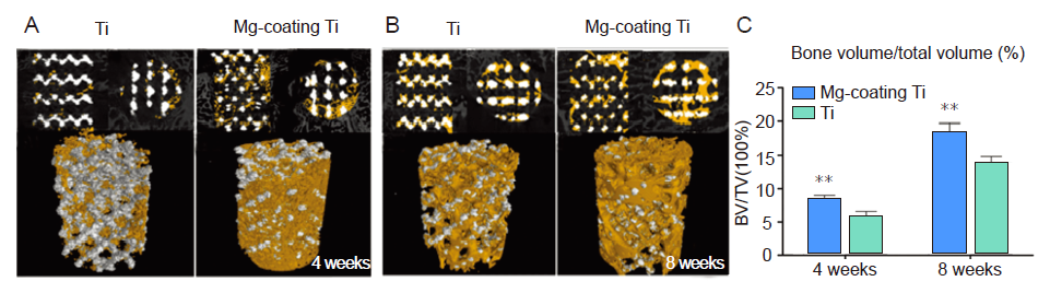
Figure 6.. (A, B) Micro-computed tomographic images of the porous Ti6Al4V with and without magnesium (Mg) coating at 4 and 8 weeks after implantation, where the yellow colour component was the newly-formed bone in these scaffolds. (C) Quantitative results showing the percentage of regenerated bone volume/total volume. Ti: titanium. *P < 0.01, vs. Ti Reprinted from Li et al.