)
)
.
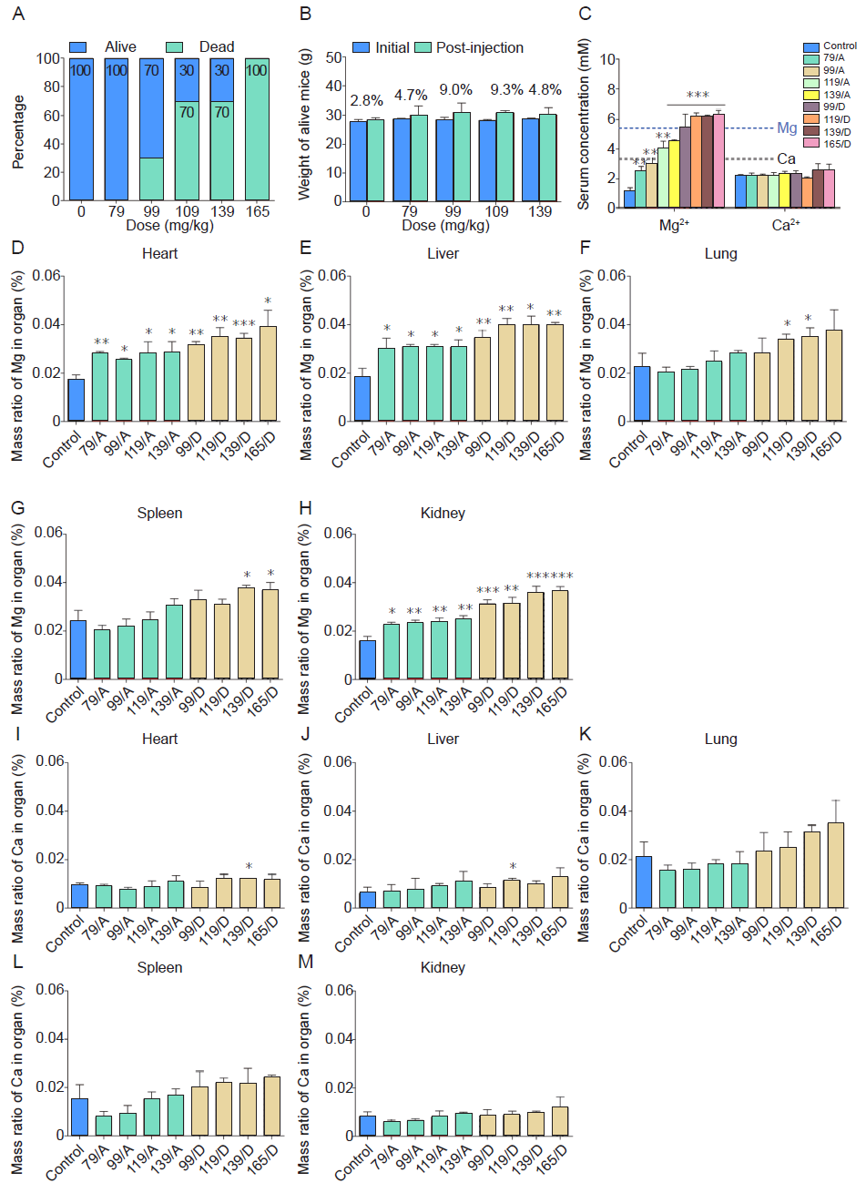
Figure 6.. In vivo median lethal dosing results of mice after intraperitoneal injection of different concentrations of diluted extract MEM. The extract MEM was prepared by soaking the PT15M scaffold in MEM at 37 °C for 72 hours. (A) The percentage of live and dead animals. For those mice that survived or dead, corresponding to the different magnesium concentrations of MEM solutions they were injected, we grouped them into nine groups which were control group (serum-free MEM), 79 mg/kg Mg/A, 99 mg/kg Mg/A, 119 mg/kg Mg/A, 119 mg/kg Mg/D, 139 mg/kg Mg/D, and 165 mg/kg Mg/D groups. (B) The weight of the live animals. The x-axis indicates Mg content in extract solution versus the weight of the mice. (C) Mg and Ca ion concentrations in the serum of mice at 1 hour after intraperitoneal injection. (D﹣H) The mass ratios of Mg in different organs. (I-M) The mass ratios of Ca in different organs. The control group was injected with MEM with a pH value of 7.0. Data are expressed as the mean ± SD (n = 10, 5 male and 5 female). *P < 0.05, **P < 0.01, ***P < 0.001 (one-way analysis of variance followed by Tukey’s post hoc test). “A” stands for alive mice, and “D” means dead mice. Ca: calcium; MEM: serum-free minimum essential medium; Mg: magnesium.