The long and winding road: homeostatic and disordered haematopoietic microenvironmental niches: a narrative review
|
The long and winding road: homeostatic and disordered haematopoietic microenvironmental niches: a narrative review |
| Suzanne M. Watt |
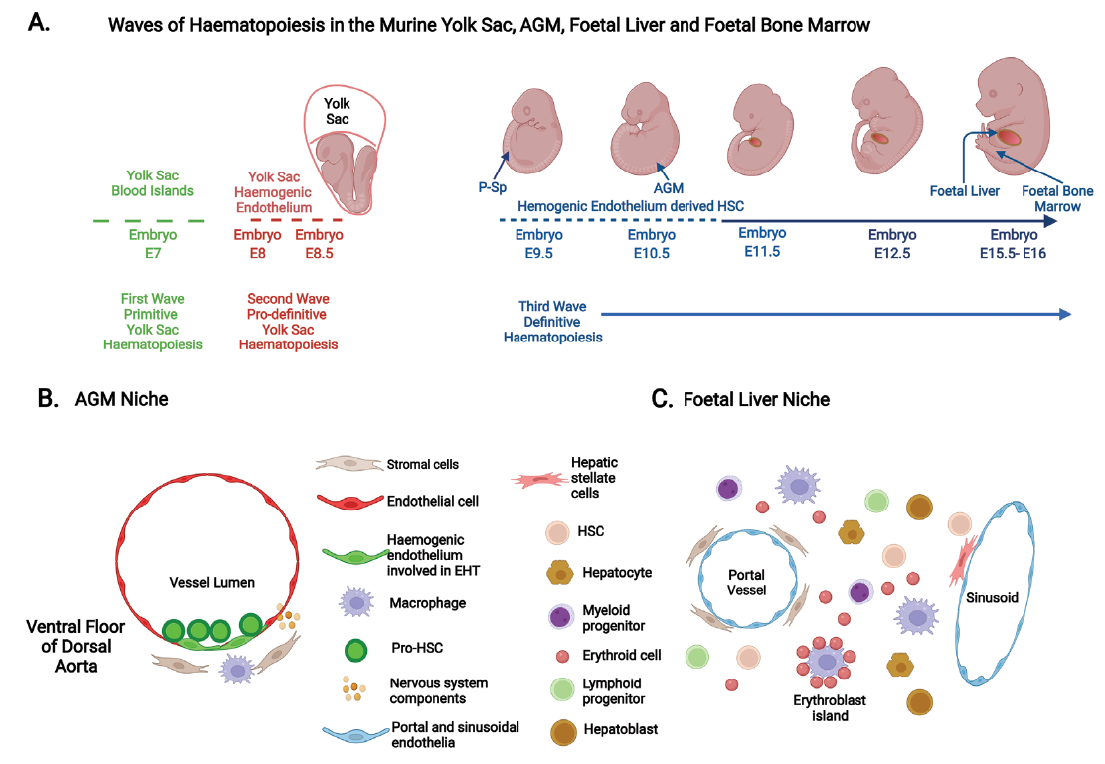
| Figure 1. Haematopoietic ontogeny in murine models. (A) The three distinct waves of haematopoiesis that occur in the developing murine embryo. The first primitive wave sees the emergence of haematopoietic cells (nucleated erythroid cells, macrophages and megakaryocytes) at E7 (7 days post-conception) from the blood islands of the yolk sac. The second pro-definitive wave arises from haemogenic endothelium of the vascular plexus of the yolk sac by the process of endothelial-haematopoietic transition (EHT) commencing at E8–8.5 and generates erythro-myeloid progenitors and certain innate immune cells. The para-aortic splanchnopleura (P-Sp) and the aorta-gonad-mesonephros (AGM) region of the embryo proper become the first and principal site of immature hematopoietic stem cell (or pro-haematopoietic stem cell (HSC)) production between E9.5–10.5. These pro-HSCs migrate (between E10.5–11) to the foetal liver, where they mature, proliferate, self-renew and/or differentiate into lymphoid and myeloid cells. The foetal liver then becomes the major haematopoietic organ until E15.5. Foetal liver HSCs migrate to the foetal bone marrow, which becomes the main residence of HSCs in adulthood. (B) A schematic cross-section of the murine embryonic AGM region with pro-HSCs emerging from the ventral floor of the dorsal aorta from haemogenic endothelia by the process of EHT. Here components of the HSC niche include endothelia, mesenchymal stromal cells, macrophages, and sympathetic nerve components. (C) A diagrammatic representation of cells present in the murine foetal liver microenvironment at E14.5 and where HSCs expand, self-renew and differentiate. These include endothelial cells of the portal and sinusoidal vessels, perivascular stromal cells, hepatic stellate cells, hepatocytes and hepatoblasts that produce cytokines, macrophages, proliferating HSCs and various haematopoietic progenitors, both myeloid and lymphoid as well as erythroid cells. Created with Biorender.com. |
|
|