Biomaterials Translational ›› 2022, Vol. 3 ›› Issue (2): 134-141.doi: 10.12336/biomatertransl.2022.02.005
• RESEARCH ARTICLE • Previous Articles Next Articles
Feifei Pu, Wei Wu, Doudou Jing, Yihan Yu, Yizhong Peng, Jianxiang Liu, Qiang Wu, Baichuan Wang, Zhicai Zhang( ), Zengwu Shao()
Received:2022-04-01
Revised:2022-04-22
Accepted:2022-05-14
Online:2022-06-28
Published:2022-06-28
Contact:
Zhicai Zhang,Zengwu Shao
E-mail:zhicaizhang@hust.edu.cn;1985XH0536@hust.edu.cn
About author:Zhicai Zhang, zhicaizhang@hust.edu.cn.
Pu, F.; Wu, W.; Jing, D.; Yu, Y.; Peng, Y.; Liu, J.; Wu, Q.; Wang, B.; Zhang, Z.; Shao, Z. Three-dimensional-printed titanium prostheses with bone trabeculae enable mechanical-biological reconstruction after resection of bone tumours. Biomater
Transl. 2022, 3(2), 134-141.

Figure 1. Image data acquisition and 3D reconstruction of tumour invasion area. (A, B) The CT scan data were imported into the medical image segmentation processing software Mimics 19.0 for image selection processing. The red arrows indicate the tumour. (C) The 3D operation to create the 3D reconstruction data of the tumour site. The blue indicates normal bone, the red indicates the bone invaded by the tumour, and the boundary between blue and purple represent the osteotomy line. B: below; L: left; T: top.
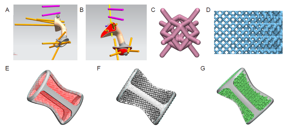
Figure 2. Design of three-dimensional printed customised prostheses. (A, B) Designing the size, fit and screw position of the prosthesis in different viewing angles. The pink and yellow sticks represent screws and connecting rods. (C) The dodecahedral lightweight lattice structure. (D) The gradient mesh designed according to the stress distribution identified by finite element analysis. (E) Design of prosthetic stiffeners and dodecahedron grid array diagram. (F) Design of gradient grid. (G) Physical design of the prosthesis.
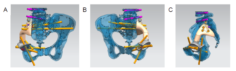
Figure 3. Computer simulation cutting and installation. (A-C) Mimics 19.0 software was used to simulate model cutting and prosthetic installation in different viewing angles.
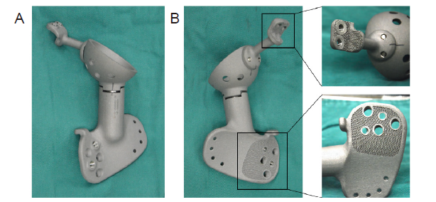
Figure 4. Preparation of three-dimensional printed personalised titanium alloy prostheses. (A, B) Titanium alloy (Ti6Al4V) was used for rapid printing by selective laser melting, and the surface interface of the prosthesis was created with a porous bone trabecular structure.
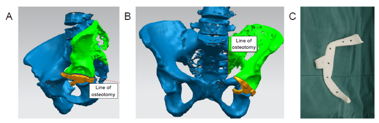
Figure 5. Three-dimensional printed navigation template design and processing. (A, B) The navigation template was designed according to the patient’s anatomical structure and surgical needs in different viewing angles. The green colour shows the part needing to be removed. (C) Preparation of the navigation template to assist with precise tumour resection.
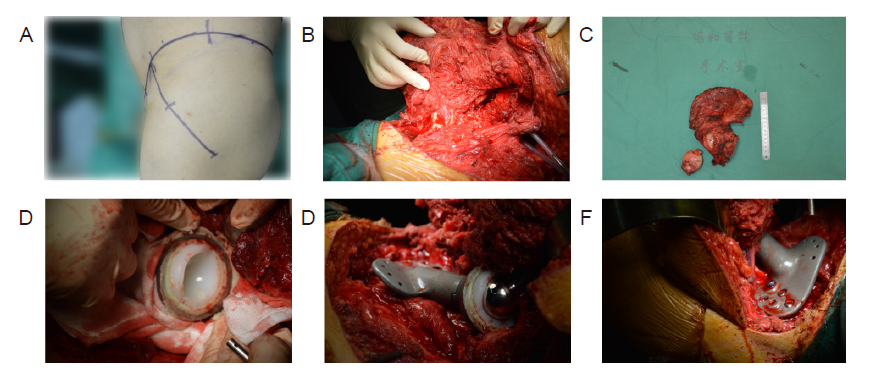
Figure 6. Implantation of a three-dimensional printed prosthesis to reconstruct a bone defect after tumour resection. (A) Surgical position and incision. (B) En bloc resection of the tumour. (C) Gross view of the excised tumour specimen. (D) Installation of the acetabular prosthesis. (E) Placing of the three-dimensional printed prosthesis in the defect. (F) The three-dimensional printed prosthesis was secured with screws.
| No. | Sex | Age (years) | Pathological type | GTV (cm3) | FU (months) | SD (minutes) | IBL (mL) | PC | TR | DM | MSTS score |
|---|---|---|---|---|---|---|---|---|---|---|---|
| 1 | Male | 42 | Ewing’s sarcoma | 320 | 16 | 375 | 1800 | No | No | No | 22 |
| 2 | Female | 37 | Malignant neurinoma | 360 | 14 | 260 | 2500 | No | No | No | 25 |
| 3 | Female | 58 | Osteosarcoma | 460 | 12 | 265 | 800 | No | No | No | 18 |
| 4 | Male | 69 | Chondrosarcoma | 300 | 23 | 270 | 2500 | No | No | No | 20 |
| 5 | Male | 58 | Chondrosarcoma | 350 | 7 | 180 | 2000 | Skin necrosis | No | No | 23 |
| 6 | Male | 42 | Osteosarcoma | 290 | 24 | 245 | 1800 | No | No | No | 20 |
| 7 | Male | 63 | Invasive GCTB | 340 | 14 | 255 | 2500 | No | No | No | 21 |
| 8 | Male | 50 | Osteosarcoma | 260 | 18 | 375 | 2000 | No | No | No | 24 |
| 9 | Male | 53 | Osteosarcoma | 280 | 16 | 280 | 2200 | No | No | No | 26 |
| 10 | Female | 48 | Chondrosarcoma | 300 | 12 | 320 | 1500 | No | No | No | 16 |
| 11 | Male | 53 | Chondrosarcoma | 260 | 18 | 260 | 2200 | No | No | No | 24 |
| 12 | Female | 46 | Chondrosarcoma | 280 | 22 | 235 | 2300 | No | No | No | 20 |
Table 1. Commonly-used gene- and cell-activated biomaterials
| No. | Sex | Age (years) | Pathological type | GTV (cm3) | FU (months) | SD (minutes) | IBL (mL) | PC | TR | DM | MSTS score |
|---|---|---|---|---|---|---|---|---|---|---|---|
| 1 | Male | 42 | Ewing’s sarcoma | 320 | 16 | 375 | 1800 | No | No | No | 22 |
| 2 | Female | 37 | Malignant neurinoma | 360 | 14 | 260 | 2500 | No | No | No | 25 |
| 3 | Female | 58 | Osteosarcoma | 460 | 12 | 265 | 800 | No | No | No | 18 |
| 4 | Male | 69 | Chondrosarcoma | 300 | 23 | 270 | 2500 | No | No | No | 20 |
| 5 | Male | 58 | Chondrosarcoma | 350 | 7 | 180 | 2000 | Skin necrosis | No | No | 23 |
| 6 | Male | 42 | Osteosarcoma | 290 | 24 | 245 | 1800 | No | No | No | 20 |
| 7 | Male | 63 | Invasive GCTB | 340 | 14 | 255 | 2500 | No | No | No | 21 |
| 8 | Male | 50 | Osteosarcoma | 260 | 18 | 375 | 2000 | No | No | No | 24 |
| 9 | Male | 53 | Osteosarcoma | 280 | 16 | 280 | 2200 | No | No | No | 26 |
| 10 | Female | 48 | Chondrosarcoma | 300 | 12 | 320 | 1500 | No | No | No | 16 |
| 11 | Male | 53 | Chondrosarcoma | 260 | 18 | 260 | 2200 | No | No | No | 24 |
| 12 | Female | 46 | Chondrosarcoma | 280 | 22 | 235 | 2300 | No | No | No | 20 |
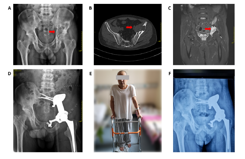
Figure 7. A 53-year-old patient with chondrosarcoma of the pelvis underwent reconstruction with a three-dimensional-printed prosthesis to repair the bone defect after tumour resection. (A-C) Preoperative X-ray images (A), computed tomography (B), and magnetic resonance imaging (C). The red arrows indicate the tumour. (D) One week postoperatively, X-ray imaging showed good position and stability of the prosthesis. (E) One week postoperatively, the patient was walking with the aid of a walker. (F) Six months after the operation, X-ray imaging showed good fusion of the prosthesis with the bone, without fracture or other complications.
| 1. |
Strauss, S. J.; Whelan, J. S. Current questions in bone sarcomas. Curr Opin Oncol. 2018, 30, 252-259.
doi: 10.1097/CCO.0000000000000456 URL |
| 2. | Zekry, K. M.; Yamamoto, N.; Hayashi, K.; Takeuchi, A.; Alkhooly, A. Z. A.; Abd-Elfattah, A. S.; Elsaid, A. N. S.; Ahmed, A. R.; Tsuchiya, H. Reconstruction of intercalary bone defect after resection of malignant bone tumor. J Orthop Surg (Hong Kong). 2019, 27, 2309499019832970. |
| 3. |
Gangi, A.; Tsoumakidou, G.; Buy, X.; Quoix, E. Quality improvement guidelines for bone tumour management. Cardiovasc Intervent Radiol. 2010, 33, 706-713.
doi: 10.1007/s00270-009-9738-9 URL |
| 4. |
Piccioli, A.; Rossi, B.; Sacchetti, F. M.; Spinelli, M. S.; Di Martino, A. Fractures in bone tumour prosthesis. Int Orthop. 2015, 39, 1981-1987.
doi: 10.1007/s00264-015-2956-7 URL |
| 5. |
Severyns, M.; Briand, S.; Waast, D.; Touchais, S.; Hamel, A.; Gouin, F. Postoperative infections after limb-sparing surgery for primary bone tumors of the pelvis: Incidence, characterization and functional impact. Surg Oncol. 2017, 26, 171-177.
doi: 10.1016/j.suronc.2017.03.005 URL |
| 6. |
Pu, F.; Liu, J.; Shi, D.; Huang, X.; Zhang, J.; Wang, B.; Wu, Q.; Zhang, Z.; Shao, Z. Reconstruction with 3D-printed prostheses after sacroiliac joint tumor resection: a retrospective case-control study. Front Oncol. 2021, 11, 764938.
doi: 10.3389/fonc.2021.764938 URL |
| 7. |
Wang, B.; Hao, Y.; Pu, F.; Jiang, W.; Shao, Z. Computer-aided designed, three dimensional-printed hemipelvic prosthesis for peri-acetabular malignant bone tumour. Int Orthop. 2018, 42, 687-694.
doi: 10.1007/s00264-017-3645-5 URL |
| 8. |
Liu, W.; Shao, Z.; Rai, S.; Hu, B.; Wu, Q.; Hu, H.; Zhang, S.; Wang, B. Three-dimensional-printed intercalary prosthesis for the reconstruction of large bone defect after joint-preserving tumor resection. J Surg Oncol. 2020, 121, 570-577.
doi: 10.1002/jso.25826 URL |
| 9. |
Hu, H.; Liu, W.; Zeng, Q.; Wang, S.; Zhang, Z.; Liu, J.; Zhang, Y.; Shao, Z.; Wang, B. The personalized shoulder reconstruction assisted by 3D printing technology after resection of the proximal humerus tumours. Cancer Manag Res. 2019, 11, 10665-10673.
doi: 10.2147/CMAR.S232051 URL |
| 10. |
Wu, J.; Xie, K.; Luo, D.; Wang, L.; Wu, W.; Yan, M.; Ai, S.; Dai, K.; Hao, Y. Three-dimensional printing-based personalized limb salvage and reconstruction treatment of pelvic tumors. J Surg Oncol. 2021, 124, 420-430.
doi: 10.1002/jso.26516 URL |
| 11. |
Bolia, I. K.; Savvidou, O. D.; Kang, H. P.; Chatzichristodoulou, N.; Megaloikonomos, P. D.; Mitsiokapa, E.; Mavrogenis, A. F.; Papagelopoulos, P. J. Cross-cultural adaptation and validation of the Musculoskeletal Tumor Society (MSTS) scoring system and Toronto Extremity Salvage Score (TESS) for musculoskeletal sarcoma patients in Greece. Eur J Orthop Surg Traumatol. 2021, 31, 1631-1638.
doi: 10.1007/s00590-021-02921-5 URL |
| 12. |
Chen, X.; Xu, L.; Wang, Y.; Hao, Y.; Wang, L. Image-guided installation of 3D-printed patient-specific implant and its application in pelvic tumor resection and reconstruction surgery. Comput Methods Programs Biomed. 2016, 125, 66-78.
doi: 10.1016/j.cmpb.2015.10.020 URL |
| 13. |
Hao, Y.; Luo, D.; Wu, J.; Wang, L.; Xie, K.; Yan, M.; Dai, K.; Hao, Y. A novel revision system for complex pelvic defects utilizing 3D-printed custom prosthesis. J Orthop Translat. 2021, 31, 102-109.
doi: 10.1016/j.jot.2021.09.006 URL |
| 14. |
Li, X.; Ji, T.; Huang, S.; Wang, C.; Zheng, Y.; Guo, W. Biomechanics study of a 3D printed sacroiliac joint fixed modular hemipelvic endoprosthesis. Clin Biomech (Bristol, Avon). 2020, 74, 87-95.
doi: 10.1016/j.clinbiomech.2020.02.014 URL |
| 15. |
Ji, T.; Yang, Y.; Tang, X.; Liang, H.; Yan, T.; Yang, R.; Guo, W. 3D-printed modular hemipelvic endoprosthetic reconstruction following periacetabular tumor resection: early results of 80 consecutive cases. J Bone Joint Surg Am. 2020, 102, 1530-1541.
doi: 10.2106/JBJS.19.01437 URL |
| 16. |
Wei, R.; Guo, W.; Ji, T.; Zhang, Y.; Liang, H. One-step reconstruction with a 3D-printed, custom-made prosthesis after total en bloc sacrectomy: a technical note. Eur Spine J. 2017, 26, 1902-1909.
doi: 10.1007/s00586-016-4871-z URL |
| 17. | Cai, H.; Liu, Z.; Wei, F.; Yu, M.; Xu, N.; Li, Z. 3D printing in spine surgery. Adv Exp Med Biol. 2018, 1093, 345-359. |
| 18. |
Xie, K.; Guo, Y.; Zhao, S.; Wang, L.; Wu, J.; Tan, J.; Yang, Y.; Wu, W.; Jiang, W.; Hao, Y. Partially melted Ti6Al4V particles increase bacterial adhesion and inhibit osteogenic activity on 3D-printed implants: an in vitro study. Clin Orthop Relat Res. 2019, 477, 2772-2782.
doi: 10.1097/CORR.0000000000000954 URL |
| 19. |
Jing, Z.; Zhang, T.; Xiu, P.; Cai, H.; Wei, Q.; Fan, D.; Lin, X.; Song, C.; Liu, Z. Functionalization of 3D-printed titanium alloy orthopedic implants: a literature review. Biomed Mater. 2020, 15, 052003.
doi: 10.1088/1748-605X/ab9078 URL |
| 20. |
Hua, L.; Lei, T.; Qian, H.; Zhang, Y.; Hu, Y.; Lei, P. 3D-printed porous tantalum: recent application in various drug delivery systems to repair hard tissue defects. Expert Opin Drug Deliv. 2021, 18, 625-634.
doi: 10.1080/17425247.2021.1860015 URL |
| 21. |
Wang, H.; Su, K.; Su, L.; Liang, P.; Ji, P.; Wang, C. Comparison of 3D-printed porous tantalum and titanium scaffolds on osteointegration and osteogenesis. Mater Sci Eng C Mater Biol Appl. 2019, 104, 109908.
doi: 10.1016/j.msec.2019.109908 URL |
| 22. |
Wei, X.; Liu, B.; Liu, G.; Yang, F.; Cao, F.; Dou, X.; Yu, W.; Wang, B.; Zheng, G.; Cheng, L.; Ma, Z.; Zhang, Y.; Yang, J.; Wang, Z.; Li, J.; Cui, D.; Wang, W.; Xie, H.; Li, L.; Zhang, F.; Lineaweaver, W. C.; Zhao, D. Mesenchymal stem cell-loaded porous tantalum integrated with biomimetic 3D collagen-based scaffold to repair large osteochondral defects in goats. Stem Cell Res Ther. 2019, 10, 72.
doi: 10.1186/s13287-019-1176-2 URL |
| 23. |
Zhang, T.; Wei, Q.; Fan, D.; Liu, X.; Li, W.; Song, C.; Tian, Y.; Cai, H.; Zheng, Y.; Liu, Z. Improved osseointegration with rhBMP-2 intraoperatively loaded in a specifically designed 3D-printed porous Ti6Al4V vertebral implant. Biomater Sci. 2020, 8, 1279-1289.
doi: 10.1039/C9BM01655D URL |
| 24. | Yin, C.; Zhang, T.; Wei, Q.; Cai, H.; Cheng, Y.; Tian, Y.; Leng, H.; Wang, C.; Feng, S.; Liu, Z. Surface treatment of 3D printed porous Ti6Al4V implants by ultraviolet photofunctionalization for improved osseointegration. Bioact Mater. 2022, 7, 26-38. |
| 25. |
Zhang, T.; Wei, Q.; Zhou, H.; Zhou, W.; Fan, D.; Lin, X.; Jing, Z.; Cai, H.; Cheng, Y.; Liu, X.; Li, W.; Song, C.; Tian, Y.; Xu, N.; Zheng, Y.; Liu, Z. Sustainable release of vancomycin from micro-arc oxidised 3D-printed porous Ti6Al4V for treating methicillin-resistant Staphylococcus aureus bone infection and enhancing osteogenesis in a rabbit tibia osteomyelitis model. Biomater Sci. 2020, 8, 3106-3115.
doi: 10.1039/C9BM01968E URL |
| 26. |
Xu, L.; Qin, H.; Cheng, Z.; Jiang, W. B.; Tan, J.; Luo, X.; Huang, W. 3D-printed personalised prostheses for bone defect repair and reconstruction following resection of metacarpal giant cell tumours. Ann Transl Med. 2021, 9, 1421.
doi: 10.21037/atm-21-3400 URL |
| 27. |
Xu, L.; Qin, H.; Tan, J.; Cheng, Z.; Luo, X.; Tan, H.; Huang, W. Clinical study of 3D printed personalized prosthesis in the treatment of bone defect after pelvic tumor resection. J Orthop Translat. 2021, 29, 163-169.
doi: 10.1016/j.jot.2021.05.007 URL |
| 28. |
Beltrami, G.; Ristori, G.; Nucci, A. M.; Galeotti, A.; Tamburini, A.; Scoccianti, G.; Campanacci, D.; Innocenti, M.; Capanna, R. Custom-made 3D-printed implants as novel approach to reconstructive surgery after oncologic resection in pediatric patients. J Clin Med. 2021, 10, 1056.
doi: 10.3390/jcm10051056 URL |
| No related articles found! |
| Viewed | ||||||
|
Full text |
|
|||||
|
Abstract |
|
|||||


