


Introduction
The significance of the built environment and the materials that construct it has become critical during the ongoing pandemic. How long does the severe acute respiratory syndrome coronavirus 2 (SARS-CoV-2) survive in the air and on surfaces? Since the pandemic hit this has been a perpetual question with the answer still being investigated. It is well known that contaminated surfaces are significant vectors in the transmission of infection both in hospitals and in the community.1, 2 Recent investigations3 into determining stability of the virus on different surfaces and in the aerosolized form revealed that the SARS-CoV-2 virus can survive for 3 hours in the aerosolized form and 72 hours on plastic and stainless steel4 with survivability varying based on indoor and outdoor conditions.5, 6 The transmission forms for both SARS-CoV-1 and SARS-CoV-2 are similar.3, 7 Studies on transmission routes for influenza virus have documented the dominant influence of airborne transmission via suspended and settled droplets8-10 resulting in guidelines of social distancing (~6 feet, about 1.83 m) to prevent person to person transmission.11
The studies all focus on obtaining data on either the influence of air circulation such as sampling in an aerosolized environment or via inoculating different types of surfaces. The investigations do not account for the fact that the two events are not isolated. Aerosolized virus laden droplets must be transported to the boundaries near the surfaces, deposit from the boundary layer on to the surface and subsequently adhere. Recent events have also shown the high probability of aerosolized transmission.12-14 The question that needs to be answered to successfully limit transmission is, “What conditions control the transport, deposition, adhesion, and persistence of airborne SARS-CoV-2 in air and on surfaces?” The interface of the boundary layer and the surface influences the transport and deposition of particles including virus laden droplets. Figure 1 illustrates the concept. The fluid mechanical boundary layer is the flow region (air) very near the surface where viscous forces dominate and transitions to a region of high velocity of air.15 Air flow in indoor spaces is often treated as well mixed but, investigations have shown the significant influence of near surface air motion on particle deposition16 and resuspension.17 There is however a lack of information on the relationship between the magnitude of shear forces and boundary flow velocity characteristics on the transport mechanism of viruses particularly near surfaces. Released virus droplets or aerosolized viruses will over time reach the boundary layers near the surface where they will subsequently ‘attach’ to the surface and adhere to it. Investigations in aqueous suspensions have shown the wide ranging and variable influence of wall shear rates on the deposition, adhesion and detachment of particles and microbes.18-20 The attachment process is influenced by the surface characteristics which include surface roughness (RH), porosity or morphology, or microbial characteristics which are mobility, flexibility, or hydrophilicity.21, 22
Figure 1.
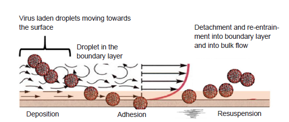
Figure 1.
Schematic illustration of the fate and transport of virus particles at the intersection of bulk and boundary layer flow.
To gain insight on these processes and their influence on the fate and transport of microbes in the indoor space, the authors assessed the adhesion behavior of a model enveloped virus, vaccinia virus (VACV), on various types of surface seen in the built environment under flow conditions. The investigation is a prelude to further studies on how the complex interaction of the multiple variables of an indoor space can influence the fate and transport of microbes in the bulk flow and boundary layer. This article also summarizes the current knowledge on particle deposition, attachment and re-entrainment in indoor spaces and the influence of properties of materials on the particle transport behavior. The paper concludes by discussing the results of the authors’ work in context of past investigations and future directions.
Methods
VACV culturing and purification
Vero cells (Centers for Disease Control and Prevention, Atlanta, GA, USA) were cultured in Dulbecco’s modified Eagle’s medium (Corning, Manassas, VA, USA) supplemented with 10% fetal bovine serum (Atlanta Biologicals, Flowery Branch, GA, USA), 2 mM L-glutamine, 1 mM sodium pyruvate, and 100 U/mL penicillin-streptomycin (HyClone, Marlborough, MA, USA). Cell cultures were maintained at 37°C in a CO2 incubator with 5% CO2 and 95% air. VACV was propagated in Vero cells with the presence of 2% fetal bovine serum Dulbecco’s modified Eagle’s medium. The infected cells were incubated at 37°C for 2 to 4 days until the microscopic cytopathic effect was complete.
The virus purification method was modified from Hruby et al.23 In brief, the infected cells were harvested and subjected to homogenization. Cell debris was removed by centrifugation at 12,296 × g for 15 minutes at 4°C (Sorvall® RC5C plus, Newtown, CT, USA). The virus pellets were precipitated by centrifugation at 81,799 × g for 3 hours at 4°C (Beckman L8-70M, Palo Alto, CA, USA) and resuspended in 1 mM Tris buffer (pH 8) overnight. Purified virus was obtained through a side-band-pull from a gradient of sucrose centrifuged at 22,504 × g for 40 minutes at 4°C. Vero cells were plated in 6-well plates at a density of 6 × 10⁵ cells per well one day prior experiment. Ten-fold serial dilutions of purified virus were made in phosphate buffered saline. The virus samples of each dilution were placed onto the prepared 6-well plate cultured with Vero cells, then incubated at room temperature in a laminar flow hood for 30 minutes. After infection, unbound virus was removed and replaced with 1% agarose (VWR Life Science, Radnor, PA, USA) in 2% fetal bovine serum Dulbecco’s modified Eagle’s medium. The plates were incubated at 37°C and 5% CO2 for 2 to 4 days until able to visualize plaques. The virus titer was calculated using an average number of plaques, dilution factor, and the inoculum volume as described elsewhere.24
Quartz crystal microbalance with dissipation analysis
The quartz crystal microbalance with dissipation (QCM-D) approach was applied to investigate virus adhesion and detachment on four types of sensor surfaces.25, 26 QCM-D technique was performed using Q-sense E4 (Biolin Scientific, Gothenburg, Sweden). Polished AT-cut 5-MHz quartz crystals coated with four materials were selected: gold (QSX 301) with RH < 1 nm, SiO2 (QSX 303) with RH < 1 nm, sodalime-glass (QSX 337) with RH < 20 nm, and stainless-steel (QSX 304) with RH < 1 nm. Temperature in the modules was controlled at 23°C. Three flow rates were assessed: 8.33 × 10⁻⁶ m3/s (50 µL/min),1.67 × 10-6 m3/s (100 µL/min), and 3.33 × 10⁻⁶ m3/s (200 µL/min).Before the experiment, all sensors were pre-cleaned with ultraviolet/ozone light for 10 minutes to remove organic contaminants on the surface. MilliQ water was first injected into the modules to provide a baseline measurement. Then, virus suspension suspended in 1 mM Tris buffer was injected at a selected flow rate. Experiments were initiated at four initial concentrations, 6.45 × 10⁴, 2.00 × 10⁵, 2.25 × 10⁵ and 2.68 × 10⁵ plaque-forming unit/mL. Virus attachment resulted in the decrease of frequency and increase of dissipation. Once both frequency and dissipation attained a constant value, the modules were rinsed with the baseline solution. Changes of frequency (∆f) and dissipation (∆D) on the sensor crystal were recorded using QSoft401 and the data were analyzed using QSense Dfind software (Biolin Scientific). Before the start of any experiment and in between each experiment, QCM-D modules and sensors were thoroughly cleaned using a 2% sodium dodecyl sulfate solution (Fisher Scientific, Waltham, MA, USA) and ultraviolet/ozone treatment and dried with N2 gas.
The QCMD detects mass and dissipation change through the excitation of the sensor crystal which oscillates at a specific frequency due to the application of a certain voltage across the electrodes because of piezoelectric properties.27, 28 Mass change (∆m) on the sensor crystal causes frequency change (∆f) and is defined by Sauerbrey relation as shown in equation (1) where c is 17.7 ng/Hz/cm2 for a 5-MHz crystal, and n is the overtone.
Dissipation (D) is caused by the adsorption of the viscoelastic film and is described as the ratio of dissipated and stored energy.29, 30 Energy lost during crystal oscillation is calculated using the measured dissipation based on equation (2) where, where Ediss is the energy dissipated during one oscillatory cycle and Estrd is the energy stored in the oscillation system.31, 32
Characterization of virus coated surfaces
Atomic force microscopy
Glass slides were cleaned by Piranha solution (30% hydrogen peroxide and concentrated sulfuric acid with 3:7 ratio from Fisher) at 75°C for 2 hours following by washed and sonicated in MilliQ water. The cleaned glass slides were then immerged in 1 mg/mL poly(diallyl dimethylammonium chloride) (PDDA) solution overnight, rinsed with MilliQ water (Millipore, Burlington, MA, USA) and dried with N2 gas. PDDA (molecular weight 100,000 to 200,000) was purchased from Sigma-Aldrich (St. Louis, MO, USA). The PDDA-glass substrate was then immerged in VACV solution at the concentration of 1 × 10⁴ plaque-forming unit/mL for 1 hour, gently rinsed with water and dried with N2 gas. Atomic force microscopy images of VACV on glass slides and RH were obtained by SPA 300 instrument (Veeco, Santa Barbara, CA, USA) in ambient conditions under tapping mode at a scan rate of 1 Hz and scan size of 10 × 10 µm2.
Fluorescence microscopy
A recombinant VACV expressing green fluorescence protein was placed onto a glass slide and evaporated to dryness in a laminar flow hood. The excessive green fluorescence protein-tagged viruses were gently washed out by phosphate buffered saline solution. The fluorescence images were acquired using a fluorescent microscope (Olympus IX81, Shinjuku-ku, Tokyo, Japan) with disk scanning unit confocal mode.
Zeta potential measurement
The purified virions were incubated for 5 minutes and overnight in 1 mM Tris buffer pH 8 and pH 4.5. The virus samples were then transferred to a DTS1070 disposable capillary cell (Malvern, Malvern, Worcestershire, UK) for zeta potential measurements. The measurements were performed at 25°C with a Zetasizer Nano-ZS (Malvern).
Transmission electron microscopy imaging
The virus was dropped onto a carbon coated copper grid for 30 minutes, and the excess virus solution was blotted off. The grid was washed with several drops of MilliQ water and dried by slow evaporation in air at room temperature. After the adsorption, 2% uranyl acetate was applied to the grid for negative staining. The morphology of the purified VACV was characterized by Transmission electron microscopy (Hitachi HT7800, Chiyoda-ku, Tokyo, Japan).
Results
VACV characteristics
VACV, a member of the poxvirus family, is an enveloped virus.33 It consists of a large double-stranded DNA genome approximately 190 kb in length, a protein layer known as the palisade, and the envelope composed of surface tubules, enveloped proteins, and lipid membranes, as illustrated in Figure 2A.34-37 VACV has an oval to brick-shaped architecture with the dimensions of 270 nm in diameter and 350 nm in length.35, 36 The purified VACV using in this study expresses green fluorescence protein and can be observed under a fluorescent microscope as shown in Figure 2B. Surface charge of the virus is one of the important factors that play an essential role in numerous sorption processes, for example adsorption and adhesion, which are governed by electrostatic interactions.38 To investigate the physical and charge characteristics of the purified virions, atomic force microscopy and dynamic light scattering were performed. In Figure 2C, VACV shows a negative surface charge with a zeta potential of approximately -30 mV in pH 8 Tris buffer solution and -20 mV in pH 4.5 Tris buffer solution, incubated in the buffers for 5 minutes and overnight, using Zetaseizer. In addition, atomic force microscopy revealed that a good deposition of negatively charged virions on a positively charged PDDA-glass substrate was found (Figure 2D). The average size of VACV is ~200-350 nm in different directions as observed under transmission electron microscope (Figure 2E), which is consistent with literature survey.39-41
Figure 2.
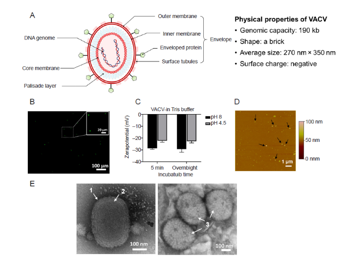
Figure 2.
VACV structural and physical properties. (A) Schematic illustration of the VACV structure, genomic capacity, shape, average size, and surface charge. (B) Fluorescence imaging of green fluorescence protein-tagged VACV (green). (C) Zeta potentials of VACV over the pH 8 and pH 4.5 in 1 mM Tris buffer. Data are expressed as mean ± SD. (D) Atomic force microscopy image of VACV deposited on PDDA-glass substrate. The black arrows point to VACV particles. (E) Representative transmission electron microscopic images of VACV. The white arrows point to (1) outer membrane, (2) core membrane, and (3) surface tubules. Scale bars: 100 μm in B, 20 µm in enlarged part in B, 1 µm in D and 100 nm in E. PDDA: poly(diallyl dimethylammonium chloride); VACV: vaccinia virus. The duplicate samples were measured and two experiments were repeated to acquire the data.
Virus adhesion kinetics via QCM-D
Table 1 shows the details of the frequency and dissipation shifts for both adhesion and detachment, and Figure 3A-D display the frequency (∆f), and dissipation (∆D) shifts of the third overtone. Four types of sensors were applied to investigate the relation between the types of sensor material and virus adhesion, gold, silica, glass, and stainless-steel (SS).
Table 1 Frequency (∆f) and dissipation (∆D) shift due to VACV adhesion and mass (∆m) of adhered VACV on the sensor surface calculated by Sauerbrey relation
| Sensor | VACV adhesion | VACV detachment | |||
|---|---|---|---|---|---|
| ∆f (Hz) | ∆D (×10-6) | ∆f (Hz) | ∆D (×10-6) | ||
| Gold | -5.77 | 1.90 | -5.37 | 1.39 | |
| SiO2 | -31.70 | 1.85 | -27.60 | 0.98 | |
| Glass | -29.81 | 2.52 | -27.22 | 1.72 | |
| Stainless-steel | -22.16 | 2.40 | -21.23 | 2.11 | |
Note: VACV: vaccinia virus. This is representative of an experiment started at 2.00 × 10⁵ plaque-forming unit/mL.
Figure 3.
Figure 3.
Quartz crystal microbalance with dissipation analysis of the frequency (∆f, black line) and dissipation (∆D, red line) (×10-6) shifts of VACV on gold (A), SiO2 (B), glass (C), and stainless-steel (D). Black arrow: VACV injection; blue arrow: rinsing with MilliQ. (E) Mass of adhered and remained virus layer on the sensor surfaces calculated by Sauerbrey relation. SS: stainless-steel; VACV: vaccinia virus. This is representative of an experiment started at 2.00 × 10⁵ plaque-forming unit/mL.
MilliQ water was first entered for a baseline. A stable baseline was observed prior to VACV injection regardless of the type of sensor material. The black arrows indicated the injection of VACV (2.0 × 10⁵ plaque-forming unit/mL). At 4 minutes, sensors were exposed to the virus suspension, thus, rapid decrease in frequency was observed due to sensor - virus bindings. Rapid increase in dissipation was observed corresponding to the virus adhesion to the sensor surface. The frequency and energy dissipation were monitored in real time while the virus adhesion resulted in the built up of multilayers on the sensor crystals. When no more significant changes in frequency and dissipation were observed, MilliQ water (blue arrows in Figure 3A-D) flow was started to rinse the sensor surface and to measure the virus detachment, concurrently. Gold sensor resulted in the smallest change of frequency, ∆f = -5.77 Hz while silica had the maximum frequency shifts of -31.70 Hz. Glass had a frequency shift of -29.81 Hz while SS exhibited a frequency shift between silica and glass, -22.16 Hz. ∆D for all sensors ranged from 1.85 × 10⁻⁶ to 2.52 × 10⁻⁶. Increase in frequency and decrease in dissipation were observed due to virus detachment and is an indicator of virus - surface adhesion characteristics i.e., whether the film layers formed is soft or rigid or became rigid due to multi-layer formation. Even though increase in frequency and decrease in dissipation were measured after rinsing, the values did not return to baseline levels. This indicates that the cleaning step washed away soft layers, but remaining layers on the sensor crystals had become rigid and could not be ‘cleaned’ thoroughly to negligible values. Figure 3E shows the calculated mass of virus layers after adhesion and then after rinsing i.e., extent of detachment applying the Sauerbrey relation. For SiO2, 13% of mass was removed after rinsing (189.1 ng/cm2 of adsorbed virus layer and 164.6 ng/cm2 of remained virus layer), followed by glass, 9% (177.8 and 162.4 ng/cm2), gold, 7% (34.4 and 32.0 ng/cm2). The lowest removal occurred for stainless-steel after rinsing (132.2 and 126.7 ng/cm2).
Figure 4 displays the f-D plots to assess the structural conformation of the adhered VACV layers. The slope (K, ∆D/∆f) represents the adsorption kinetic process on the sensor crystal. Higher K indicates the structural conformation of the layer on the sensor surface is soft and flexible whereas lower K represents that the layer is thin and rigid. Initially a soft layer (K1) is formed while as it adheres to the sensor surface and then the layer becomes firm (K2). After the rinsing step, part of the adhered layer has been removed from the sensor (increase in frequency) and the remaining layer is more rigid (K3).
Figure 4.

Figure 4.
Frequency-dissipation plots for VACV for gold (A), SiO2 (B), glass (C), and stainless-steel (D). ‘1’, ‘2’, and ‘3’ show the steps of the adhesion process, ‘1’ adhesion, ‘2’ reaching saturation and ‘3’ detachment due to the wash cycle. The numbers correspond to the slope represented by K1, K2 and K3. VACV: vaccinia virus. This is representative of an experiment started at 2.00 × 10⁵ plaque-forming unit/mL.
Figure 4A shows the adsorption process for VACV-gold. In comparison to Figure 4B-D which represents the adhesion process of VACV for silica, glass, and SS respectively, K1 and K2 is the steepest for gold sensor indicating the layer attached is more viscoelastic compared to the others. The sensor also has the lowest K3 value which indicates that the layer remaining is thin, and rigid as opposed to its counterparts. Figure 4B and C show similar characteristics of the layer formed initially at the beginning of adherence of VACV to silica and glass. K2 and K3 values for silica are less than glass. VACV-SS interaction based on K1, K2 and K3 is very similar in magnitude to glass. SS surface is positively charged42 and the extent of detachment of negatively charged VACV is significantly less compared to the other sensors.
Discussion
The results focus on one aspect of the multi-faceted problem - influence of surface type on attachment and detachment of VACV. The significance of understanding how surface properties control the adhesion kinetics is highlighted. During the initial adhesion process the viscoelastic nature of the virus layer was dependent on the surface type. After the first phase of attachment, there is a plateau indicating saturation has been reached and the layer is becoming rigid. Rinse cycle is started immediately, and the extent washed off differs based on the properties of the layer formed and for these surface types, surface charge. Gold behaves very differently from the other surfaces, in that while mass attached is lower than SS, it remains attached. Glass and silica appear to be the ‘cleanest’ surfaces since adhered viruses were easily cleaned compared to other surfaces. The effect of ‘flow’ on the behavior of VACV-surface interactions remains to be investigated and correlated. In the sections below the factors that can influence fate and transport of aerosols including microbes are discussed.
Phenomena influencing deposition
Extensive investigation in the factors influencing deposition of inorganic particles has been done because of the significant role it plays on human health and exposure. The deposition phenomena are influenced by multiple factors which include particle characteristics, air flow, interior design, and surface coverings.43-46 The interacting effect of ventilation, location of furniture and air changes have shown that while higher air changes removed particles faster, localized exposure and deposition is influenced by a combination of multiple factors.47 Experimental and modeling studies determined a lumped parameter: deposition velocity or loss rate coefficient for a range of particle sizes to distinguish the effects of the multi-dimensional design space which describe the indoor environment.48, 49 Deposition velocities and loss rate coefficients provide a bulk perspective of the transport and removal of particles from the air.50 The rate at which deposition occurred is represented by deposition velocity, vd as shown in $v_{d}=\frac{M}{t C A_{s}}$ where M is the mass of particle on a sample surface, t is time of exposure, C is time-weighted average mass concentration of particles in air and As is the surface sample area.
Studies in small scale chambers and real houses have shown that particle removal by deposition is significantly correlated to diameter, surface to air temperature difference, surface orientation, spatial location, and RH.43, 46, 49 Deposition constants have been shown to be related to building wall textures, orientation and particle size.51 Among surface properties, the influence of RH has been assessed. Lai and Nazaroff52 observed that particle deposition increased for most particle sizes onto smooth and rough vertical surfaces, with roughness simulated using smooth glass plates and sandpaper. While deposition clearly increased with near wall airflow velocity the influence of RH became less evident48 probably due to the dominance of the fluid momentum boundary layer.
Deposition velocities in relation to microbe carrying particles or for microbes have not been investigated extensively. Typically, the studies incorporate properties or stay within the range of parameters accepted for inorganic particles. For example, a computational fluid dynamics model based on Eulerian-Lagrangian framework simulated the deposition of Staphylococcus and Micrococcus, the conclusions identified that mixing and ventilation conditions influenced deposition of the bacterial species.53 The spherical species have a diameter ~1 μm and were selected because of the proximity of the physical characteristics for the application of stokes’ law and lack of information for microbes. Studies cannot account for the characteristics of the different types of microbes, bacteria, viruses, and fungi which can influence transport and deposition. Whyte and Eaton54 calculated deposition velocities as a function of concentration of airborne particles carrying microbes by collecting samples from clean rooms and operation theatres. They reported higher deposition rates for lower concentrations. Seong and Hoque55 assessed the influence of sampling region and sampling location on bacterial species detected.
Adhesion forces governing surface interaction
The forces encountered in adhesion of solid particles on solid surfaces either in air (at different humidity levels) or in water or other media are molecular interactions defined by Van der Waals’ forces, electrostatic interaction, liquid bridges, double layer interaction and polar and/or metallic bonds.56-59 Dust and activated carbon appear to preferentially adhere to insulated surfaces such as polyvinyl chloride or glass compared to aluminum and copper.60 Physics-based model such as the Hamaker model depending exclusively on Van der Waals forces was not successful in interpreting the adhesion mechanism and results indicated the significance of considering polar contributions.60, 61 Other investigations have looked into particle shape and size pointing out the limitation that most studies tend to focus on spherical particles.62, 63
Deryaguin-Muller-Toporov, Johnson-Kendall-Roberts and Maugis-Pollock models64-66 have been applied to describe molecular attraction forces and the influence of contact areas between particles and surfaces. RH and contact angle have a high impact on the magnitude of the Van der Waals forces. Higher humidity levels, beyond 50% tend to enhance adhesion;56, 61 however, the hydrophilic/hydrophobic nature of particles and surfaces impacts the degree of influence.67 For example, adhesion force of glass on glass or glass on silica surfaces for diameters ~20 to 60 μm treated to be hydrophobic remained constant for all humidity levels while for hydrophilic conditions, at 50-60% humidity adhesion force increases or resuspension decreased.68 The anomaly observed for hydrophobic surfaces and the increasing deviation from classical theoretical predictions for larger size particles have been attributed to the water film formed between particles and surfaces and/or the electrostatic forces. Adhesion force magnitudes decreasing with particle size.56 Particles of size in the range > 1 μm to ~5 μm adherence to surfaces is determined by the nature of the contact and are harder to resuspend but larger size particles are easier to resuspend and the adhesion mechanism is influenced by contact points, and geometry.57, 69
This literature survey on adhesion of microbiological particles to surfaces target bacteria and viruses. Studies focusing on bacteria are dominated by areas such as biocorrosion,70 biomaterial implants,71, 72 environmental microbiology,73 food industry,74 and microbial fuel cells.75 The adhesion mechanism is modeled typically using the Derjaguin, Landau, Verwey, Overbeek (DLVO) approach, or the thermodynamic approach or the extended DLVO model.76-79 The DLVO theory is based on the non-specific interaction energies between the van der Waals forces and the electrostatic double layer forces which can be attractive or repulsive contingent on the bacteria-surface combination. Thermodynamic theory on the other hand is based on the concept of surface free energies which would account for the various types of interactions including van der Waals, electrostatic and dipole moments. Since the DLVO theory assumed inert chemical surfaces, a modification was added to the theory by adding a short-range Lewis acid-base term which will account for the hydrophobicity/hydrophilicity in an extended DLVO theory.80
The extended DLVO theory has been applied to shed light on the mechanisms governing the adhesion of viruses to certain surfaces. For example, a study by Chrysikopoulos and Syngouna81 looked at the interaction of bacteriophages, MS2 and ΦX174 with clay colloids. The virus attachment was described by the Freundlich isotherm and the Lewis acid-base term in the extended DLVO model was critical in explaining the hydrophobic interaction mediated attachment. The extended DLVO approach was utilized to model the attachment of human adenoviruses and two bacteriophages, P22 and MS282 to lip balms. The study showed that drying of the lip balms resulted in the drop of surface free energy which made the surfaces highly hydrophobic. The extended DLVO model results predicted that attachment was favored due to short range strong hydrophobic interaction. Hydrophobic and electrostatic interactions were also shown to govern the attachment of MS2 bacteriophage to surfaces treated with polyelectrolyte multilayers.83
Recent investigations have tried to unravel the implications of virus adhesion kinetics with regard to health effects, infection transmission and SARS-CoV-24,84-86 by utilizing representative surrogates such as a lentivirus85 or through conducting a theoretical analysis utilizing existing data to assess the influence of different surfaces and environmental conditions including temperature, humidity, and pH.6, 84, 87 Experimental methods for determining the adhesion force and kinetic mechanisms include using the centrifuge approach88, 89 or the QCM-D82, 83, 90 and AFM.91, 92 Liu et al.93 used floor dust as surrogates for fungal spore and high-speed imaging to capture the effect of velocity on resuspension. For microbes and surfaces of the built environment, investigation has focused on antimicrobial properties of metal alloys such as copper - zinc, copper - silver on surfaces and their application.94-97 The studies highlight the significant differences that exist between the adhesion mechanisms for inorganic particles versus microbes.98
The mechanism of resuspension
Measurements of resuspension have been expressed as resuspension factors or resuspension rates.99 Resuspension factor is the ratio of air borne contaminant concentration per unit air volume to the contaminant surface concentration per unit area on the ground and resuspension rate is defined as the fraction of a surface species removed in unit time.99
Experimental results showed that smaller particles required larger flow velocity to achieve the same amount of detachment as the larger particles. The detachment fraction was also dependent on the surface adhesion energy of the particles ranging between ~32 μm and ~76 μm with higher velocities required for higher adhesion energy.100 Punjrath and Heldman101 studied particle entrainment in a wind tunnel and theorized that two mechanisms: a) initiation of particle movement when shear stresses on the particles exceed friction forces acting on the particles and b) transfer of momentum from other moving particles dominated resuspension.
Modeling studies comprised of two approaches - statistical and/or force balance. Theoretical models have simulated the mechanisms at a micro scale,17, 102-104 whereas computational fluid dynamics models have focused on capturing the movement of a specific activity such as foot tapping62 and analytical models105 have applied multiple fundamental concepts such as dimensional analysis to model the effects. The Eulerian method, in which particles are treated as a continuum, has been traditionally applied to the cases of heavy particle deposits while Lagrangian methods have been applied to individually track relatively light particles in monolayer or few multi-layer systems.106, 107 In Lagrangian-based models, particle transport is generally modeled through the addition of gravitational, drag, added mass, Saffman’s lift, bed impact forces and Bassett forces.108 Among these forces, Bassett forces (force due to acceleration of the particle) have been considered negligible and bed impact forces are more dominant than lift forces. Braaten et al.102 assumed that fluid forces are applied at the surface in discrete ‘bursts’ following a probability distribution. Saffman’s lift force109 has been used in the literature to describe the particle motion when studying particle deposition mainly. Mollinger and Nieuwstadt110 and Leighton and Acrivos111 also developed expressions for the lift force for particles touching the wall. Shi and Bayless112 developed a computational fluid dynamics model of a gas-particle flow in cyclones. The authors incorporated a balance of adhesion and lift-off forces to account for particle detachment from the surfaces.
Due to the inherent random nature of particle behavior, statistical approaches for predicting resuspension such as Monte Carlo simulations113 and Lagrangian stochastic models have been proposed.114 The stochastic models showed that resuspension can be captured numerically but appropriate attention needs to be given to fluctuations created due to turbulent flow or bursts created due to surface impacts.17, 103 Loosmore99 developed empirical models to calculate resuspension rate, using five parameters: friction velocity, time since the wind flow begin, particle diameter, particle density, and roughness height and identified friction velocity and time of exposure as the most important factors. In an indoor space particle resuspension magnitude could vary by two to three orders of magnitude. Resuspension rates between 3 × 10⁻⁷ and 6 × 10⁻⁶ min-1 were found for super micron particles of density 1000 kg/m3. Substantial resuspension of particles of diameter 2.5 μm and 5 μm occurred with source strengths ranging from 0.03 mg/min to 0.5 mg/min, a range estimated for human activities.43, 44, 115
Few studies have explored the effects of resuspension of microbes. Krauter and Biermann116 examined the reaerosolization of dry spores (0.6-1.1 μm) in a ventilation duct. Resuspension rates of fungal spores on both steep and plastic duct materials were between 6 × 10⁻² and 6 × 10⁻⁴ min-1, which decreased to 10 times less than the initial rates within 30 minutes. In depth analysis of the influence of friction, RH, exposure time and forces which have been assessed to an extent for particles have not been quantified regarding bacteria, viruses, or fungi.
Conclusions
The current pandemic has reinforced the necessity of establishing baseline information on how viruses under indoor environmental conditions optimize survivability and transmission. Based on the discussions above, investigations into understanding this phenomenon can be separated into three main categories. One category investigates the effects of indoor environmental factors and surface properties on boundary flow characteristics. The second group of studies assesses the influence of the same variables on various phenomena such as drag force, electrostatic force or thermophoretic force which effects aerosol fate and transport. The last category is the effect of physical and biological properties on the deposition, attachment, and persistence of microbes on surfaces. Typically, the three categories discussed are investigated independent of each other with some overlap occurring in categories 1 and 2. But, an indoor environment with a range of bacteria, viruses and fungal species, and an evolving microbial world encompasses all three categories as shown in Figure 5. To successfully understand, assess, and predict how the indoor environment perpetuates transmission, we must bridge the gaps between these fields.
Figure 5.
Figure 5.
A schematic representation of the current state and future research directions. f: function.
Author contributions
Study concepts: SH and QW; study design: SH, DS, MK, YL and QW; previous studies analysis and manuscript review: SH, DS, MK and QW; experiment implementation and data analysis: DS, MK and YL; data acquisition: DS and MK; manuscript preparation: SH, DS and MK; manuscript editing: SH, DS, MK and YL. All authors reviewed and approved the final version of the manuscript.
Financial support
We would like to thank the Office of the Vice President of Research, University of South Carolina for the funds under COVID-19 research grants. Yuan Lin would like to thank the support from the Chinese Scholarship Council (No. 201904910172).
Acknowledgement
None.
Conflicts of interest statement
Qian Wang is an Editorial Board member of Biomaterials Translational.
Data sharing statement
This is an open access journal, and articles are distributed under the terms of the Creative Commons Attribution-NonCommercial-ShareAlike 4.0 License, which allows others to remix, tweak, and build upon the work non-commercially, as long as appropriate credit is given and the new creations are licensed under the identical terms.
Reference
Possible transmission by fomites of respiratory syncytial virus
DOI:10.1093/infdis/141.1.98
URL
PMID:7365274
[Cited within: 1]

To test whether nosocomial spread of respiratory syncytial virus (RSV) could occur through contact with environmental surfaces contaminated by RSV-infected nasal secretions, survival in the environment of RSV isolated from media, pooled adult secretions, and secretions from hospitalized infants was examined. RSV in freshly obtained infant secretions was recovered from countertops for up to 6 hr, from rubber gloves for up to 1 1/2 hr, from cloth gowns and paper tissue for 30--45 min, and from skin for up to 20 min. RSV in media and pooled secretions survived for slightly longer periods. Further experiments demonstrated that infectious virus could be transferred to hands touching these contaminated surfaces and could be recovered from these hands for up to 25 min. These studies suggest that survival of RSV in the environment of infected infant secretions is sufficient to allow transfer of infectious virus to the hands of hospital personnel. Thus, self-inoculation by contact with contaminated infant secretions may be a potential mode of nosocomial transmission of RSV.
The effects of temperature and relative humidity on the viability of the SARS coronavirus
DOI:10.1155/2011/734690
URL
PMID:22312351
[Cited within: 1]

The main route of transmission of SARS CoV infection is presumed to be respiratory droplets. However the virus is also detectable in other body fluids and excreta. The stability of the virus at different temperatures and relative humidity on smooth surfaces were studied. The dried virus on smooth surfaces retained its viability for over 5 days at temperatures of 22-25 degrees C and relative humidity of 40-50%, that is, typical air-conditioned environments. However, virus viability was rapidly lost (>3 log(10)) at higher temperatures and higher relative humidity (e.g., 38 degrees C, and relative humidity of >95%). The better stability of SARS coronavirus at low temperature and low humidity environment may facilitate its transmission in community in subtropical area (such as Hong Kong) during the spring and in air-conditioned environments. It may also explain why some Asian countries in tropical area (such as Malaysia, Indonesia or Thailand) with high temperature and high relative humidity environment did not have major community outbreaks of SARS.
Aerosol and surface stability of SARS-CoV-2 as compared with SARS-CoV-1
Stability of SARS-CoV-2 in different environmental conditions
DOI:10.1016/S2666-5247(20)30003-3 URL PMID:32835322 [Cited within: 2]
Environmental factors involved in SARS-CoV-2 transmission: effect and role of indoor environmental quality in the strategy for COVID-19 infection control
DOI:10.1186/s12199-020-00904-2
URL
PMID:33143660
[Cited within: 1]
The severe acute respiratory syndrome coronavirus 2 (SARS-CoV-2), a new zoonotic agent that emerged in December 2019, causes coronavirus disease 2019 (COVID-19). This infection can be spread by asymptomatic, presymptomatic, and symptomatic carriers. SARS-CoV-2 spreads primarily via respiratory droplets during close person-to-person contact in a closed space, especially a building. This article summarizes the environmental factors involved in SARS-CoV-2 transmission, including a strategy to prevent SARS-CoV-2 transmission in a building environment. SARS-CoV-2 can persist on surfaces of fomites for at least 3 days depending on the conditions. If SARS-CoV-2 is aerosolized intentionally, it is stable for at least several hours. SARS-CoV-2 is inactivated rapidly on surfaces with sunlight. Close-contact aerosol transmission through smaller aerosolized particles is likely to be combined with respiratory droplets and contact transmission in a confined, crowded, and poorly ventilated indoor environment, as suggested by some cluster cases. Although evidence of the effect of aerosol transmission is limited and uncertainty remains, adequate preventive measures to control indoor environmental quality are required, based on a precautionary approach, because COVID-19 has caused serious global damages to public health, community, and the social economy. The expert panel for COVID-19 in Japan has focused on the
Increasing temperature and relative humidity accelerates inactivation of SARS-CoV-2 on surfaces
DOI:10.1128/mSphere.00441-20
URL
PMID:32611701
[Cited within: 2]
Coronavirus disease 2019 (COVID-19) was first identified in China in late 2019 and is caused by newly identified severe acute respiratory syndrome coronavirus 2 (SARS-CoV-2). Previous studies had reported the stability of SARS-CoV-2 in cell culture media and deposited onto surfaces under a limited set of environmental conditions. Here, we broadly investigated the effects of relative humidity, temperature, and droplet size on the stability of SARS-CoV-2 in a simulated clinically relevant matrix dried on nonporous surfaces. The results show that SARS-CoV-2 decayed more rapidly when either humidity or temperature was increased but that droplet volume (1 to 50 mul) and surface type (stainless steel, plastic, or nitrile glove) did not significantly impact decay rate. At room temperature (24 degrees C), virus half-life ranged from 6.3 to 18.6 h depending on the relative humidity but was reduced to 1.0 to 8.9 h when the temperature was increased to 35 degrees C. These findings suggest that a potential for fomite transmission may persist for hours to days in indoor environments and have implications for assessment of the risk posed by surface contamination in indoor environments.IMPORTANCE Mitigating the transmission of SARS-CoV-2 in clinical settings and public spaces is critically important to reduce the number of COVID-19 cases while effective vaccines and therapeutics are under development. SARS-CoV-2 transmission is thought to primarily occur through direct person-to-person transfer of infectious respiratory droplets or through aerosol-generating medical procedures. However, contact with contaminated surfaces may also play a significant role. In this context, understanding the factors contributing to SARS-CoV-2 persistence on surfaces will enable a more accurate estimation of the risk of contact transmission and inform mitigation strategies. To this end, we have developed a simple mathematical model that can be used to estimate virus decay on nonporous surfaces under a range of conditions and which may be utilized operationally to identify indoor environments in which the virus is most persistent.
SARS in hospital emergency room
DOI:10.3201/eid1005.030579
URL
PMID:15200809
[Cited within: 1]
Thirty-one cases of severe acute respiratory syndrome (SARS) occurred after exposure in the emergency room at the National Taiwan University Hospital. The index patient was linked to an outbreak at a nearby municipal hospital. Three clusters were identified over a 3-week period. The first cluster (5 patients) and the second cluster (14 patients) occurred among patients, family members, and nursing aids. The third cluster (12 patients) occurred exclusively among healthcare workers. Six healthcare workers had close contact with SARS patients. Six others, with different working patterns, indicated that they did not have contact with a SARS patient. Environmental surveys found 9 of 119 samples of inanimate objects to be positive for SARS coronavirus RNA. These observations indicate that although transmission by direct contact with known SARS patients was responsible for most cases, environmental contamination with the SARS coronavirus may have lead to infection among healthcare workers without documented contact with known hospitalized SARS patients.
Dynamics of infectious disease transmission by inhalable respiratory droplets
DOI:10.1098/rsif.2010.0026
URL
PMID:20164087
[Cited within: 1]
Transmission of respiratory infectious diseases in humans, for instance influenza, occurs by several modes. Respiratory droplets provide a vector of transmission of an infectious pathogen that may contribute to different transmission modes. An epidemiological model incorporating the dynamics of inhalable respiratory droplets is developed to assess their relevance in the infectious process. Inhalable respiratory droplets are divided into respirable droplets, with droplet diameter less than 10 microm, and inspirable droplets, with diameter in the range 10-100 microm: both droplet classes may be inhaled or settle. Droplet dynamics is determined by their physical properties (size), whereas population dynamics is determined by, among other parameters, the pathogen infectivity and the host contact rates. Three model influenza epidemic scenarios, mediated by different airborne or settled droplet classes, are analysed. The scenarios are distinguished by the characteristic times associated with breathing at contact and with hand-to-face contact. The scenarios suggest that airborne transmission, mediated by respirable droplets, provides the dominant transmission mode in middle and long-term epidemics, whereas inspirable droplets, be they airborne or settled, characterize short-term epidemics with high attack rates. The model neglects close-contact transmission by droplet sprays (direct projection onto facial mucous membranes), retaining close-contact transmission by inspirable droplets.
Airborne spread of infectious agents in the indoor environment
DOI:10.1016/j.ajic.2016.06.003
URL
PMID:27590694
BACKGROUND: Since the 2003 severe acute respiratory syndrome epidemic, scientific exploration of infection control is no longer restricted to microbiologists or medical scientists. Many studies have reported on the release, transport, and exposure of expiratory droplets because of respiratory activities. This review focuses on the airborne spread of infectious agents from mucus to mucus in the indoor environment and their spread as governed by airflows in the respiratory system, around people, and in buildings at different transport stages. METHODS: We critically review the literature on the release of respiratory droplets, their transport and dispersion in the indoor environment, and the ultimate exposure of a susceptible host, as influenced by airflows. RESULTS: These droplets or droplet nuclei are transported by expired airflows, which are sometimes affected by the human body plume and use of a face mask, as well as room airflow. Room airflow is affected by human activities such as walking and door opening, and some droplets are eventually captured by a susceptible individual because of his or her inspired flows; such exposure can eventually lead to long-range spread of airborne pathogens. Direct exposure to the expired fine droplets or droplet nuclei results in short-range airborne transmission. Deposition of droplets and direct personal exposure to expired large droplets can lead to the fomite route and the droplet-borne route, respectively. CONCLUSIONS: We have shown the opportunities for infection control at different stages of the spread. We propose that the short-range airborne route may be important in close contact, and its control may be achieved by face masks for the source patients and use of personalized ventilation. Our discussion of the effect of thermal stratification and expiratory delivery of droplets leads to the suggestion that displacement ventilation may not be applicable to hospital rooms where respiratory infection is a concern.
Short-range airborne transmission of expiratory droplets between two people
DOI:10.1111/ina.12314
URL
PMID:27287598
[Cited within: 1]
The occurrence of close proximity infection for many respiratory diseases is often cited as evidence of large droplet and/or close contact transmission. We explored interpersonal exposure of exhaled droplets and droplet nuclei of two standing thermal manikins as affected by distance, humidity, ventilation, and breathing mode. Under the specific set of conditions studied, we found a substantial increase in airborne exposure to droplet nuclei exhaled by the source manikin when a susceptible manikin is within about 1.5 m of the source manikin, referred to as the proximity effect. The threshold distance of about 1.5 m distinguishes the two basic transmission processes of droplets and droplet nuclei, that is, short-range modes and the long-range airborne route. The short-range modes include both the conventional large droplet route and the newly defined short-range airborne transmission. We thus reveal that transmission occurring in close proximity to the source patient includes both droplet-borne (large droplet) and short-range airborne routes, in addition to the direct deposition of large droplets on other body surfaces. The mechanisms of the droplet-borne and short-range airborne routes are different; their effective control methods also differ. Neither the current droplet precautions nor dilution ventilation prevents short-range airborne transmission, so new control methods are needed.
Public health guidance for potential COVID-19 exposure associated with travel
https://www.cdc.gov/coronavirus/2019-ncov/php/risk-assessment.html. Accessed by March 30,
The COVID-19 pandemic in the US: a clinical update
URL PMID:32250388 [Cited within: 1]
A choir decided to go ahead with rehearsal. Now dozens of members have COVID-19 and two are dead
Airborne route and bad use of ventilation systems as non-negligible factors in SARS-CoV-2 transmission
URL PMID:32361528 [Cited within: 1]
Indoor boundary layer chemistry modeling
DOI:10.1111/ina.12601
URL
PMID:31461792
[Cited within: 1]
Ozone (O3 ) chemistry is thought to dominate the oxidation of indoor surfaces. We consider the hypothesis that reactions taking place within indoor boundary layers result in greater than anticipated hydroxyl radical (OH) deposition rates. We develop models that account for boundary layer mass-transfer phenomena, O3 -terpene chemistry and OH formation, removal, and deposition; we solve these analytically and by applying numerical methods. For an O3 -limonene system, we find that OH flux to a surface with an O3 reaction probability of 10(-8) is 4.3 x 10(-5) molec/(cm(2) s) which is about 10 times greater than predicted by a traditional boundary layer theory. At very low air exchange rates the OH surface flux can be as much as 10% of that for O3 . This effect becomes less pronounced for more O3 -reactive surfaces. Turbulence intensity does not strongly influence the OH concentration gradient except for surfaces with an O3 reaction probability >10(-4) . Although the O3 flux dominates OH flux under most conditions, OH flux can be responsible for as much as 10% of total oxidant uptake to otherwise low-reactivity surfaces. Further, OH chemistry differs from that for ozone; therefore, its deposition is important in understanding the chemical evolution of some indoor surfaces and surface films.
Modeling indoor particle deposition from turbulent flow onto smooth surfaces
Effects of shear on particle motion near a surface—application to resuspension
Mechanisms responsible for sub-micron particle deposition in a laminar wall-jet
Deposition of oral streptococci and polystyrene latices onto glass in a parallel plate flow cell
Microbial adhesion in flow displacement systems
DOI:10.1128/CMR.19.1.127-141.2006
URL
PMID:16418527
[Cited within: 1]
Flow displacement systems are superior to many other (static) systems for studying microbial adhesion to surfaces because mass transport and prevailing shear conditions can be adequately controlled and notoriously ill-defined slight rinsing steps to remove so-called
Escherichia coli, Pseudomonas aeruginosa, and Staphylococcus aureus attachment patterns on glass surfaces with nanoscale roughness
DOI:10.1007/s00284-008-9320-8
URL
PMID:19020934
[Cited within: 1]
Attachment tendencies of Escherichia coli K12, Pseudomonas aeruginosa ATCC 9027, and Staphylococcus aureus CIP 68.5 onto glass surfaces of different degrees of nanometer-scale roughness have been studied. Contact-angle and surface-charge measurements, atomic force microscopy (AFM), scanning electron microscopy (SEM), and confocal laser scanning microscopy (CLSM) were employed to characterize substrata and bacterial surfaces. Modification of the glass surface resulted in nanometer-scale changes in the surface topography, whereas the physicochemical characteristics of the surfaces remained almost constant. AFM analysis indicated that the overall surface roughness parameters were reduced by 60-70%. SEM, CLSM, and AFM analysis clearly demonstrates that although E. coli, P. aeruginosa and S. aureus present significantly different patterns of attachment, all of the species exhibited a greater propensity for adhesion to the
Deposition method, relative humidity, and surface property effects of bacterial spore reaerosolization via pulsed air jet
Vaccinia virus replication. I. Requirement for the host-cell nucleus
DOI:10.1128/JVI.29.2.705-715.1979
URL
PMID:107327
[Cited within: 1]
Using cytochalasin B-induced enucleation techniques, we examined the ability of vaccinia virus to replicate in the absence of the host-cell nucleus in several mammalian cell lines. It was found that virus-infected enucleated cells (cytoplasts) prepared from BSC-40, CVC, and L cells were incapable of producing infectious progeny virus. The nature of this apparent nuclear involvement was studied in detail in BSC-40 cells. Modulations designed to maximize cytoplast integrity and longevity, such as reduction of the growth temperature and initial multiplicity of infection, did not improve virus growth in cytoplasts. Sodium dodecyl sulfate-polyacrylamide gel analysis of the [(35)S]methionine pulse-labeled proteins synthesized in vaccinia virus-infected cytoplasts demonstrated that both early and late viral gene products were being expressed at high levels and with the proper temporal sequence. Vaccinia virus cytoplasmic DNA synthesis, as measured by [(3)H]thymidine incorporation, peaked at 3 h postinfection and was 70 to 90% of control levels in cytoplasts. However, in the cytoplasts this DNA was not converted to a DNase-resistant form late in infection, which was consistent with the failure to isolate physical particles from infected cytoplasts. Treatment of vaccinia virus-infected cells with 100 mug of rifampin/ml from 0 to 8 h to increase the pools of viral precursors, followed by subsequent removal of the drug, resulted in a threefold increase virus yield. This treatment had no effect on virus-infected cytoplasts. Finally, vaccinia virus morphogenesis was studied under an electron microscope in thin sections of virus-infected cells and cytoplasts which had been prepared at various times during a single-step growth cycle. It was apparent that, although early virus morphogenetic forms appeared, there was no subsequent DNA condensation or particle maturation in the cytoplasts. These results suggest that vaccinia virus requires some factor or function from the host-cell nucleus in order to mature properly and produce infectious progeny virus.
Two detailed plaque assay protocols for the quantification of infectious SARS-CoV-2
DOI:10.1002/cpmc.105
URL
PMID:32475066
[Cited within: 1]
Severe acute respiratory syndrome coronavirus-2 (SARS-CoV-2) has been identified as the causal agent of COronaVIrus Disease-19 (COVID-19), an atypical pneumonia-like syndrome that emerged in December 2019. While SARS-CoV-2 titers can be measured by detection of viral nucleic acid, this method is unable to quantitate infectious virions. Measurement of infectious SARS-CoV-2 can be achieved by tissue culture infectious dose-50 (TCID50 ), which detects the presence or absence of cytopathic effect in cells infected with serial dilutions of a virus specimen. However, this method only provides a qualitative infectious virus titer. Plaque assays are a quantitative method of measuring infectious SARS-CoV-2 by quantifying the plaques formed in cell culture upon infection with serial dilutions of a virus specimen. As such, plaque assays remain the gold standard in quantifying concentrations of replication-competent lytic virions. Here, we describe two detailed plaque assay protocols to quantify infectious SARS-CoV-2 using different overlay and staining methods. Both methods have several advantages and disadvantages, which can be considered when choosing the procedure best suited for each laboratory. These assays can be used for several research purposes, including titration of virus stocks produced from infected cell supernatant and, with further optimization, quantification of SARS-CoV-2 in specimens collected from infected animals. (c) 2019 The Authors. Basic Protocol: SARS-CoV-2 plaque assay using a solid double overlay method Alternate Protocol: SARS-CoV-2 plaque assay using a liquid overlay and fixation-staining method.
Real-time detection of airborne viruses on a mass-sensitive device
DOI:10.1063/1.2956679
URL
PMID:19529841
[Cited within: 1]
We present real-time detection of airborne Vaccinia viruses using quartz crystal microbalance (QCM) in an integrated manner. Vaccinia viruses were aerosolized and neutralized using an electrospray aerosol generator, transported into the QCM chamber, and captured by a QCM crystal. The capture of the viruses on the QCM crystal resulted in frequency shifts proportional to the number of viruses. The capture rate varied linearly with the concentration of initial virus suspensions (8.5x10(8)-8.5x10(10) particlesml) at flow rates of 2.0 and 1.1 lmin. This work demonstrates the general potential of mass sensitive detection of nanoscale biological entities in air.
The detection of influenza A and B viruses in clinical specimens using a quartz crystal microbalance
DOI:10.1016/j.jviromet.2009.07.001
URL
PMID:19628008
[Cited within: 1]
Current methods for the accurate diagnosis of influenza based on culture of the virus or PCR are highly sensitive and specific but require specialised laboratory facilities and highly trained personnel and, in the case of viral culture, can take up to 14 days to obtain a definitive result. In this study, a quartz crystal microbalance-based immunosensor (QCM) has been developed and its potential evaluated for the rapid and sensitive detection of both influenza A and B viruses in laboratory-cultured preparations and clinical samples. The effective limit for detection by QCM for stock preparations of both A/PR/8/34 and B/Lee/40 viruses was 1 x 10(4) pfu/mL, associated with observed frequency shifts of 30 (+/-5) and 37 (+/-6.5) Hz, respectively. Conjugation of 13 nm gold nanoparticles to the detecting antibody improved the mass sensitivity of the immunosensor, resulting in a 10-fold increase in sensitivity and a detection limit of 1 x 10(3) pfu/mL for both preparations, with resulting frequency shifts of 102 (+/-11) and 115 (+/-5) Hz, respectively. Detection of virus in nasal washes with this technique was achieved by overnight passage in MDCK cultures prior to analysis. A comparison of results obtained from 67 clinical samples using existing RT-PCR, shell vial, cell culture and ELISA methods showed that QCM techniques were comparable in sensitivity and specificity to cell culture methods.
Quartz crystal microbalance with dissipation monitoring (QCM-D): real-time characterization of nano-scale interactions at surfaces
Energy dissipation kinetics for protein and antibody-antigen adsorption under shear oscillation on a quartz crystal microbalance
QCM-D sensitivity to protein adsorption reversibility
DOI:10.1002/bit.21977
URL
PMID:18623227
[Cited within: 1]
Using a quartz crystal microbalance with dissipative monitoring (QCM-D) we have determined the adsorption reversibility and viscoelastic properties of ribonuclease A adsorbed to hydrophobic self-assembled monolayers. Consistent with previous work with proteins unfolding on hydrophobic surfaces, high protein solution concentrations, reduced adsorption times, and low ammonium sulfate concentrations lead to increased adsorption reversibility. Measured rigidity of the protein layers normalized for adsorbed protein amounts, a quantity we term specific dissipation, correlated with adsorption reversibility of ribonuclease A. These results suggest that specific dissipation may be correlated with changes in structure of adsorbed proteins.
Quartz crystal microbalance with dissipation monitoring: enabling real-time characterization of biological materials and their interactions
URL
PMID:19137101
[Cited within: 1]
In recent years, there has been a rapid growth in the number of scientific reports in which the quartz crystal microbalance (QCM) technique has played a key role in elucidating various aspects of biological materials and their interactions. This article illustrates some key advances in the development of a special variation of this technique called quartz crystal microbalance with dissipation monitoring (QCM-D). The main feature and advantage of QCM-D, compared with the conventional QCM, is that it in addition to measuring changes in resonant frequency (Deltaf), a simultaneous parameter related to the energy loss or dissipation (DeltaD) of the system is also measured. Deltaf essentially measures changes in the mass attached to the sensor surface, while DeltaD measures properties related to the viscoelastic properties of the adlayer. Thus, QCM-D measures two totally independent properties of the adlayer. The focus of this review is an overview of the QCM-D technology and highlights of recent applications. Specifically, recent applications dealing with DNA, proteins, lipids, and cells will be detailed. This is not intended as a comprehensive review of all possible applications of the QCM-D technology, but rather a glimpse into a few highlighted application areas in the biomolecular field that were published in 2007.
Quartz crystal microbalance with dissipation (QCM-D) studies of the viscoelastic response from a continuously growing grafted polyelectrolyte layer
DOI:10.1016/j.jcis.2013.07.008
URL
PMID:23932084
[Cited within: 1]
Poly(acrylic acid) was grown from substrates by photopolymerization, and the grafting process was monitored in situ by Quartz Crystal Microbalance with Dissipation (QCM-D) measurements in a 1:1 v/v mixture of water/ethanol. The polymerization process was monitored into the thick film region, where the change in frequency and dissipation with increasing film mass changes sign as predicted by the Voigt viscoelastic model. Our experimental data are compared with predictions of this model, and satisfactory agreement is found for low overtone numbers. The Voigt model was applied to analyze the measured changes in frequency, Deltaf, and dissipation, DeltaD, in order to extract information on layer thickness, shear elasticity, mu, and shear viscosity, eta, of the growing film. The increasing rate of changes in Deltaf and DeltaD observed after about 150s of polymerization was found to correlate with an increasing growth rate of the film thickness. For longer polymerization times a close to linear increase in thickness with time was observed. The sensitivity, defined as the derivatives of Deltaf and DeltaD with respect to thickness, depends on overtone number and is different for the frequency and dissipation signals - facts that should be considered when investigating small changes in thick films used in e.g. sensor applications.
Viscoelastic properties of fibrinogen adsorbed onto poly(ethylene terephthalate) surfaces by QCM-D
DOI:10.1016/j.carbpol.2012.02.075
URL
PMID:23465926
[Cited within: 1]
In presented study a new approach using QCM-D for biocompatibility determination was introduced. The adsorption of fibrinogen on PET and modified PET surfaces was monitored in situ using QCM-D. Protein layer thicknesses were estimated on the basis of a Voight based viscoelastic model. The hydrophilicities and morphologies of the surfaces were investigated using a goniometer and AFM. The results showed that PET surfaces coated with sulphated polysaccharides are more hydrophilic and more fibrinogen-repulsive than non-modified PET surfaces. QCM-D equipped with QTools modelling software is well-applicable to the characterisation of surface properties and can be optimised for biocompatibility determination.
Vaccinia virus morphogenesis and dissemination
DOI:10.1016/j.tim.2008.07.009
URL
PMID:18789694
[Cited within: 1]
Vaccinia virus is the smallpox vaccine. It is the most intensively studied poxvirus, and its study has provided important insights about virus replication in general and the interactions of viruses with the host cell and immune system. Here, the entry, morphogenesis and dissemination of vaccinia virus are considered. These processes are complicated by the existence of two infectious vaccinia virus particles, called intracellular mature virus (IMV) and extracellular enveloped virus (EEV). The IMV particle is surrounded by one membrane, and the EEV particle comprises an IMV particle enclosed within a second lipid membrane containing several viral antigens. Consequently, these virions have different biological properties and play different roles in the virus life cycle.
Oncolytic viruses: a new class of immunotherapy drugs
DOI:10.1038/nrd4663
URL
PMID:26323545
[Cited within: 1]
Oncolytic viruses represent a new class of therapeutic agents that promote anti-tumour responses through a dual mechanism of action that is dependent on selective tumour cell killing and the induction of systemic anti-tumour immunity. The molecular and cellular mechanisms of action are not fully elucidated but are likely to depend on viral replication within transformed cells, induction of primary cell death, interaction with tumour cell antiviral elements and initiation of innate and adaptive anti-tumour immunity. A variety of native and genetically modified viruses have been developed as oncolytic agents, and the approval of the first oncolytic virus by the US Food and Drug Administration (FDA) is anticipated in the near future. This Review provides a comprehensive overview of the basic biology supporting oncolytic viruses as cancer therapeutic agents, describes oncolytic viruses in advanced clinical trials and discusses the unique challenges in the development of oncolytic viruses as a new class of drugs for the treatment of cancer.
Vaccinia virus-mediated cancer immunotherapy: cancer vaccines and oncolytics
DOI:10.1186/s40425-018-0495-7
URL
PMID:30626434
[Cited within: 1]
Cancer vaccines and oncolytic immunotherapy are promising treatment strategies with potential to provide greater clinical benefit to patients with advanced-stage cancer. In particular, recombinant vaccinia viruses (VV) hold great promise as interventional agents. In this article, we first summarize the current understanding of virus biology and viral genes involved in host-virus interactions to further improve the utility of these agents in therapeutic applications. We then discuss recent findings from basic and clinical studies using VV as cancer vaccines and oncolytic immunotherapies. Despite encouraging results gleaned from translational studies in animal models, clinical trials implementing VV vectors alone as cancer vaccines have yielded largely disappointing results. However, the combination of VV vaccines with alternate forms of standard therapies has resulted in superior clinical efficacy. For instance, combination regimens using TG4010 (MVA-MUC1-IL2) with first-line chemotherapy in advanced-stage non-small cell lung cancer or combining PANVAC with docetaxel in the setting of metastatic breast cancer have clearly provided enhanced clinical benefits to patients. Another novel cancer vaccine approach is to stimulate anti-tumor immunity via STING activation in Batf3-dependent dendritic cells (DC) through the use of replication-attenuated VV vectors. Oncolytic VVs have now been engineered for improved safety and superior therapeutic efficacy by arming them with immune-stimulatory genes or pro-apoptotic molecules to facilitate tumor immunogenic cell death, leading to enhanced DC-mediated cross-priming of T cells recognizing tumor antigens, including neoantigens. Encouraging translational and early phase clinical results with Pexa-Vec have matured into an ongoing global phase III trial for patients with hepatocellular carcinoma. Combinatorial approaches, most notably those using immune checkpoint blockade, have produced exciting pre-clinical results and warrant the development of innovative clinical studies. Finally, we discuss major hurdles that remain in the field and offer some perspectives regarding the development of next generation VV vectors for use as cancer therapeutics.
Structure of intracellular mature vaccinia virus observed by cryoelectron microscopy
DOI:10.1128/JVI.68.3.1935-1941.1994
URL
PMID:8107253
[Cited within: 1]
Intracellular mature vaccinia virus, also called intracellular naked virus, and its core envelope have been observed in their native, unfixed, unstained, hydrated states by cryoelectron microscopy of vitrified samples. The virion appears as a smooth rounded rectangle of ca. 350 by 270 nm. The core seems homogeneous and is surrounded by a 30-nm-thick surface domain delimited by membranes. We show that surface tubules and most likely also the characteristic dumbbell-shaped core with the lateral bodies which are generally observed in negatively stained or conventionally embedded samples are preparation artifacts.
In a nutshell: structure and assembly of the vaccinia virion
DOI:10.1016/S0065-3527(06)66002-8
URL
PMID:16877059
[Cited within: 1]
Poxviruses comprise a large family of viruses characterized by a large, linear dsDNA genome, a cytoplasmic site of replication and a complex virion morphology. The most notorious member of the poxvirus family is variola, the causative agent of smallpox. The laboratory prototype virus used for the study of poxviruses is vaccinia, the virus that was used as a live, naturally attenuated vaccine for the eradication of smallpox. Both the morphogenesis and structure of poxvirus virions are unique among viruses. Poxvirus virions apparently lack any of the symmetry features common to other viruses such as helical or icosahedral capsids or nucleocapsids. Instead poxvirus virions appear as
Isoelectric points of viruses
DOI:10.1111/j.1365-2672.2010.04663.x
URL
PMID:20102425
[Cited within: 1]
Efficient removal of viruses by a novel polyvinylidene fluoride membrane filter
DOI:10.1016/s0166-0934(96)02163-5
URL
PMID:9128859
[Cited within: 1]
Virus removal by a novel filter (Ultipor VF DV50), comprising three layers of PVDF membrane, has been evaluated by infectivity studies using a range of viruses and conditions. The filter was able to remove at least 6 log of various viruses, i.e. Sindbis and Semliki Forest (40-70 nm), herpes simplex (120-200 nm) and vaccinia (200 x 350 nm), from cell-culture medium or phosphate buffered saline pH 6.8 containing 0.5% albumin. However, the removal of polio virus (25-30 nm) under these conditions was only limited, i.e. about 1 log. This filter is thus effective for removing viruses of about 50 nm or larger. Proteins as large as immunoglobulins (MW 160,000), were able to pass through the filter with recoveries of at least 85%. Due to its ability to remove viruses of medium to large size, this filter shows potential for increasing the safety of biological products where viruses such as hepatitis B, C, herpes and retroviruses are of concern.
Cryo-electron tomography of vaccinia virus
DOI:10.1073/pnas.0409825102
URL
PMID:15699328
The combination of cryo-microscopy and electron tomographic reconstruction has allowed us to determine the structure of one of the more complex viruses, intracellular mature vaccinia virus, at a resolution of 4-6 nm. The tomographic reconstruction allows us to dissect the different structural components of the viral particle, avoiding projection artifacts derived from previous microscopic observations. A surface-rendering representation revealed brick-shaped viral particles with slightly rounded edges and dimensions of approximately 360 x 270 x 250 nm. The outer layer was consistent with a lipid membrane (5-6 nm thick), below which usually two lateral bodies were found, built up by a heterogeneous material without apparent ordering or repetitive features. The internal core presented an inner cavity with electron dense coils of presumptive DNA-protein complexes, together with areas of very low density. The core was surrounded by two layers comprising an overall thickness of approximately 18-19 nm; the inner layer was consistent with a lipid membrane. The outer layer was discontinuous, formed by a periodic palisade built by the side interaction of T-shaped protein spikes that were anchored in the lower membrane and were arranged into small hexagonal crystallites. It was possible to detect a few pore-like structures that communicated the inner side of the core with the region outside the layer built by the T-shaped spike palisade.
Contact transmission of vaccinia to an infant diagnosed by viral culture and metagenomic sequencing
DOI:10.1093/ofid/ofaa111
URL
PMID:32685604
[Cited within: 1]
Targeted molecular diagnostic tests and accurate immunoassays have transformed the landscape of clinical virology, calling into question the usefulness of traditional viral culture. Here we present a case where viral culture, followed by metagenomic sequencing, was central to the diagnosis of an unexpected viral infection, with significant clinical and public health implications.
Cleanability of titanium and stainless steel particles in relation to surface charge aspects
DOI:10.4265/bio.13.9
URL
PMID:18432111
[Cited within: 1]
The cleanability of titanium and 316L stainless steel particles was studied in terms of their apparent surface charge density (sigma(app)). Bovine serum albumin (BSA) was used as the model fouling agent. Curves for the sigma(app) of titanium and stainless steel particles showed the apparent points of zero charge (pzc(app)) of 4.6 and 8.5, respectively. Compared with the curve for the sigma(app) of stainless steel, that of titanium was characterized by small positive and large negative sigma(app) values. The isotherms for BSA adsorption and the saturation amount of BSA adsorbed on titanium and stainless steel depended largely on the intrinsic properties of BSA. In continuous cleaning in a plug-flow column fed by a 0.05M NaOH solution, BSA was found to be faster desorbed from titanium than from stainless steel, and smaller amounts of BSA remaining after 120-min cleaning were observed on titanium. Kinetic analysis showed that the two first-order desorption rate constants, reflecting the rate of BSA desorption in the initial and later stages of cleaning, for titanium were respectively 1.7-fold and 1.3-fold higher than those values for stainless steel. It could be suggested that the better cleanability of titanium was probably due to the small binding strength of BSA on slightly negatively-charged titanium surfaces and due to their large negative sigma(app) values under alkaline cleaning conditions.
Particle deposition from natural convection enclosure flow onto smooth surfaces
Deposition, resuspension, and penetration of particles within a residence
Particle deposition indoors: a review
DOI:10.1034/j.1600-0668.2002.01159.x
URL
PMID:12532752
Particle deposition indoors has received increasing attention recently because of increasing concern about the effects of particle exposure on human health. Deposition is a positive phenomenon from the perspective of human health, as deposited particles cannot be inhaled unless resuspended. There are studies showing strong correlation between the prevalence of biological-origin airborne particles and the prevalence of some specific sick building syndrome (Menzies et al., 1998; Teeuw et al., 1994). In the literature, the amount of research effort put on particle deposition indoors which is directly related to human health and microcontamination control is far less than the amount of research on deposition in small diameter tube/channel. This is a summary of the full-length version which will appear on the home page. The full-length paper aims to provide an up-to-date revision for both experiment and modeling on particle deposition indoors. This paper summarizes the experimental studies for particle deposition indoors for non-industrial environments. In section 2, 'Background', an overview of the different mechanisms of particle deposition, focusing on indoor environments is addressed. In section 3, 'Experimental Study Review', a survey of aerosol deposition experiments in small experimental chambers and real houses (or large-scale chambers) is presented. Detailed experimental measurements are addressed for the real house studies. The experimental techniques of particle generation, particle labeling and detection methods are discussed. Although the present paper focuses on experimental studies, it would be more complete to include some discussions on the modeling of the term particle eddy diffusivity and it is reviewed in section 4, 'Modeling Review'.
Particle deposition in indoor environments: analysis of influencing factors
DOI:10.1016/j.jhazmat.2007.01.032
URL
PMID:17280778
[Cited within: 2]
In this paper, several factors influencing particle deposition in indoor environments are analyzed with an analytical model and a three-dimensional drift flux model combined with the particle deposition boundary conditions for wall surfaces. The influences of flow conditions near the wall surfaces, surface roughness and particle concentration distribution on particle deposition indoors are studied. By modeling particle deposition onto surfaces with the analytical model, it is found that larger friction velocity near the wall surfaces and rougher surface may lead to larger particle deposition velocity when the particle size is small, but when particle size is large enough (the range is up to the actual friction velocity and in this study it is about 1-5 microm), the influence of the friction velocity and roughness could be neglected. Furthermore, the three-dimensional numerical simulations indicate that particle concentration distribution may be very different even for the same particle source and air change rate, which cause a different deposited particle flux. As the particle concentration distribution may not be uniform in most cases, especially for the ventilated rooms, it is important to incorporate particle concentration distribution when analyzing particle deposition in indoor environments. Some suggestions or rules for particle deposition controlling are also presented based on the analysis.
Coupling computational fluid dynamics simulations and statistical moments for designing healthy indoor spaces
Deposition of fine particles on building internal surfaces
Effects of surface properties of vertical textiles indoors on particle deposition: a small-scale chamber study
Critique of the use of deposition velocity in modeling indoor air quality
Indoor particle pollution: effect of wall textures on particle deposition
Supermicron particle deposition from turbulent chamber flow onto smooth and rough vertical surfaces
An Experimental and Numerical Study on Deposition of Bioaerosols in a Scaled Chamber
Deposition velocities of airborne microbe-carrying particles
Does the presence of certain bacterial family in the microbiome indicate specific indoor environment characteristics? A factorial design approach for identifying bio-fingerprints
Adhesion and removal of fine particles on surfaces
The adhesion of solid particles to solid surfaces. I. A review
The adhesion of solid particles to solid surfaces. II
DOI:10.1080/00022470.1961.10468039 URL PMID:13881367
Particle adhesion and removal: a review
Adhesion of dust particles to common indoor surfaces in an air-conditioned environment
Adhesion and surface energy of mica in air and water
Human induced flow field and resultant particle resuspension and transport during gait cycle
Effect of geometry on particle adhesion
Adhesion between highly rough alumina surfaces: an atomic force microscope study
DOI:10.1016/j.jcis.2008.11.050
URL
PMID:19101675
[Cited within: 1]
The removal of contaminant particles in microelectronics processes now extends not only to the compounds themselves but also to the reactor pieces where they are fabricated. This raises new issues as both the particles and the reactor walls are highly rough and a maximum number of particles per unit area is tolerated. In this work we study the adhesion force of a sapphire particle onto alumina substrates of roughness ranging from 10 nm to 3 microm peak-to-peak on a 5 microm x 5 microm area, in water. A contribution of this work is the prediction of the statistic of the adhesion force by a fast running numerical computation, without any adjustable parameters. The perspective is to be able to predict the best conditions for removing contaminant particles.
Effect of contact deformations on the adhesion of particles
A model for particle removal from surfaces with large-scale roughness in turbulent flows
Effects of relative humidity and particle and surface properties on particle resuspension rates
Adhesion forces between glass and silicon surfaces in air studied by afm: effects of relative humidity, particle size, roughness, and surface treatment
Adhesion as an interplay between particle size and surface roughness
DOI:10.1016/j.jcis.2006.09.015
URL
PMID:17010358
[Cited within: 1]
Surface roughness plays an important role in the adhesion of small particles. In this paper we have investigated adhesion as a geometrical effect taking into account both the particle size and the size of the surface features. Adhesion is studied using blunt model particles on surfaces up to 10 nm root-mean-square (RMS) roughness. Measurements with particles both smaller and larger than surface features are presented. Results indicate different behavior in these areas. Adhesion of particles smaller than or similar in size to the asperities depend mainly on the size and shape of the asperities and only weakly on the size of the particle. For large particles also the particle size has a significant effect on the adhesion. A new model, which takes the relative size of particles and asperities into account, is also derived and compared to the experimental data. The proposed model predicts adhesion well over a wide range of particle/asperity length scales.
Metal surface characteristics dictate bacterial adhesion capacity
Concise review of mechanisms of bacterial adhesion to biomaterial surfaces
DOI:10.1002/(sici)1097-4636(199823)43:3<338::aid-jbm16>3.0.co;2-b
URL
PMID:9730073
[Cited within: 1]
This article reviews the mechanisms of bacterial adhesion to biomaterial surfaces and the factors affecting the adhesion. The process of bacterial adhesion includes an initial physicochemical interaction phase (phase one) and a late molecular and cellular interaction phase (phase two), which is a complicated process affected by many factors, including the characteristics of the bacteria themselves, the target material surface, and the environmental factors, such as the presence of serum proteins or bactericidal substances.
Comparison of surface roughness and bacterial adhesion between cosmetic contact lenses and conventional contact lenses
DOI:10.1097/ICL.0000000000000054
URL
PMID:25536530
[Cited within: 1]
OBJECTIVE: To compare physical characteristics of cosmetic contact lenses (Cos-CLs) and conventional contact lenses (Con-CLs) that might affect susceptibility to bacterial adhesion on the contact lens (CL) surface. METHODS: Surface characteristics of Cos-CLs and Con-CLs made from the same material by the same manufacturer were measured by atomic force microscopy (AFM) and scanning electron microscopy. To determine the extent and rate of bacterial adhesion, Cos-CL and Con-CL were immersed in serum-free Roswell Park Memorial Institute media containing Staphylococcus aureus or Pseudomonas aeruginosa. Additionally, the rate of removal of adherent bacteria was evaluated using hand rubbing or immersion in multipurpose disinfecting solutions (MPDS). RESULTS: The mean surface roughness (root mean square and peak-to-valley value) measured by AFM was significantly higher for Cos-CL than for Con-CL. At each time point, significantly more S. aureus and P. aeruginosa adhered to Cos-CL than to Con-CL, which correlated with the surface roughness of CL. In Cos-CL, bacteria were mainly found on the tinted surface rather than on the noncolored or convex areas. Pseudomonas aeruginosa attached earlier than S. aureus to all types of CL. However, P. aeruginosa was more easily removed from the surface of CL than S. aureus by hand rubbing or MPDS soaking. CONCLUSIONS: Increased surface roughness is an important physical factor for bacterial adhesion in Cos-CL, which may explain why rates of bacterial keratitis rates are higher in Cos-CL users in CL physical characteristics.
Bacterial adhesion at the single-cell level
DOI:10.1038/s41579-018-0057-5
URL
PMID:30008468
[Cited within: 1]
The formation of multicellular microbial communities, called biofilms, starts from the adhesion of a few planktonic cells to the surface. The transition from a free-living planktonic lifestyle to a sessile, attached state is a multifactorial process that is determined by biological, chemical and physical properties of the environment, the surface and the bacterial cell. The initial weak, reversible interactions between a bacterium and a surface strengthen to yield irreversible adhesion. In this Review, we summarize our understanding of the mechanisms governing bacterial adhesion at the single-cell level, including the physical forces experienced by a cell before reaching the surface, the first contact with a surface and the transition from reversible to permanent adhesion.
The adhesion and detachment of bacteria and spores on food-contact surfaces
Microbial fuel cells--challenges and applications
Numerical modeling of bacterium-surface interaction by applying DEM
Concise review of mechanisms of bacterial adhesion to biomaterials and of techniques used in estimating bacteria-material interactions
DOI:10.22203/ecm.v008a05
URL
PMID:15593018
This article reviews the mechanisms of bacterial adhesion to biomaterial surfaces, the factors affecting the adhesion, the techniques used in estimating bacteria-material interactions and the models that have been developed in order to predict adhesion. The process of bacterial adhesion includes an initial physicochemical interaction phase and a late molecular and cellular one. It is a complicated process influenced by many factors, including the bacterial properties, the material surface characteristics, the environmental factors, such as the presence of serum proteins and the associated flow conditions. Two categories of techniques used in estimating bacteria-material interactions are described: those that utilize fluid flowing against the adhered bacteria and counting the percentage of bacteria that detach, and those that manipulate single bacteria in various configurations which lend themselves to more specific force application and provide the basis for theoretical analysis of the receptor-ligand interactions. The theories that are reviewed are the Derjaguin-Landau-Verwey-Overbeek (DLVO) theory, the thermodynamic approach and the extended DLVO theory. Over the years, significant work has been done to investigate the process of bacterial adhesion to biomaterial surfaces, however a lot of questions still remain unanswered.
Quantitatively predicting bacterial adhesion using surface free energy determined with a spectrophotometric method
Assessing bacterial adhesion using DLVO and XDLVO theories and the jet impingement technique
DOI:10.1016/j.colsurfb.2009.04.030
URL
PMID:19493661
[Cited within: 1]
In this study, the adhesion of two bacterial strains (Pseudomonas stutzeri PS, and Staphylococcus epidermidis, SE) to the glass and the indium tin oxide (ITO)-coated glass surfaces was examined qualitatively and quantitatively using the theoretical approaches and the jet impingement technique. A comparison between the DLVO and the extended DLVO (XDLVO) theories showed that the XDLVO predictions of bacterial adhesion and its reversibility are more accurate than DLVO predictions. The adhesion tests revealed that PS bacteria has much better adhesion rate than SE bacteria to both material surfaces, as predicted by XDLVO approach. Also both bacterial strains adhered better to the hydrophobic ITO-coated glass than to the hydrophilic glass surface, as predicted theoretically. Moreover, the microjet impingement technique was used not only to assess the bacterial adhesion strength on both materials, but also to verify the adhesion reversibility. The detachment stress values demonstrated that PS bacterial cells adhered strongly and irreversibly in the primary energy minimum, while SE bacterial cells adhered weakly and reversibly in the secondary energy minimum on both substrata surfaces. Also, the adhesion of both bacterial strains was found better and stronger on the hydrophobic ITO-coated glass surface than on the hydrophilic glass surface.
Hydrophobicity of biosurfaces — Origin, quantitative determination and interaction energies
Attachment of bacteriophages MS2 and ΦX174 onto kaolinite and montmorillonite: extended-DLVO interactions
DOI:10.1016/j.colsurfb.2011.11.028
URL
PMID:22153836
[Cited within: 1]
This study aims to gain insights into the interaction of virus particles with clay colloids. Bacteriophages MS2 and PhiX174 were used as model viruses and kaolinite (KGa-1b) and montmorillonite (STx-1b) as model colloids. The experimental data obtained from batch experiments of MS2 and PhiX174 attachment onto KGa-1b and STx-1b suggested that virus attachment is adequately described by the Freundlich isotherm equation. Both MS2 and PhiX174 were attached in greater amounts onto KGa-1b than STx-1b with MS2 having greater affinity than PhiX174 for both clays. Furthermore, extended-DLVO interaction energy calculations explained that the attachment of viruses onto model clay colloids was primarily caused by hydrophobic interaction. The theoretical and experimental results of this study were found to be in good agreement with previous findings.
Lip balm drying promotes virus attachment: Characterization of lip balm coatings and XDLVO modeling
DOI:10.1016/j.jcis.2020.07.143
URL
PMID:32877879
[Cited within: 2]
HYPOTHESIS: Drying-induced decrease in lip balm surface energy enhances virus adhesion due to the emergence of strong hydrophobic colloid-surface interactions. EXPERIMENTS: A protocol was developed for preparing lip balm coatings to enable physicochemical characterization and adhesion studies. Surface charge and hydrophobicity of four brands of lip balm (dry and hydrated) and human adenovirus 5 (HAdV5) were measured and used to calculate the extended Derjaguin-Landau-Verwey-Overbeek (XDLVO) energy of interactions between lip balm coatings and HAdV5 as well as four other colloids: HAdV40, MS2 and P22 bacteriophages, and SiO2. Quartz crystal microbalance with dissipation monitoring (QCM-D) tests employed SiO2 colloids, HAdV5 and hydrated lip balms. FINDINGS: Drying of lip balms results in a dramatic decrease of their surface energy (deltaDeltaGsws>/= 83.0mJ/m(2)) making the surfaces highly hydrophobic. For dry lip balms, the interaction of the balm surface with all five colloids is attractive. For lip balms hydrated in 150mM NaCl (ionic strength of human saliva), XDLVO calculations predict that hydrophilic colloids (MS2, P22, SiO2) may attach into shallow secondary minima. Due to the relative hydrophobicity of human adenoviruses, primary maxima in XDLVO profiles are low or non-existent making irreversible deposition into primary energy minima possible. Preliminary QCM-D tests with SiO2 colloids and HAdV5 confirm deposition on hydrated lip balms.
Virus deposition onto polyelectrolyte-coated surfaces: A study with bacteriophage MS2
DOI:10.1016/j.jcis.2018.12.107
URL
PMID:30639663
[Cited within: 2]
HYPOTHESES: By selecting constituent polyelectrolytes and controlling conditions of their deposition, the resulting polyelectrolyte multilayers can be designed as surface coatings with controlled adhesive properties with respect to viruses. Charge and hydrophilicity of the polyelectrolyte multilayers govern virus adhesion. EXPERIMENTS: Four surfaces of different charges and hydrophobicities were designed using a layer-by-layer assembly of poly(styrene-4-sulfonate) and poly(dimethyl diallyl ammonium chloride). Contact angle measurements gave an estimate of MS2 hydrophilicity in terms of free energy of interfacial interaction in water. Experimental results on MS2 adhesion obtained using quartz crystal microbalance with dissipation monitoring were compared with predictions by the extended Derjaguin-Landau-Verwey-Overbeek (XDLVO) theory. FINDINGS: MS2 deposition onto polyelectrolyte multilayers occurred in two phases: an early phase defined by virus-surface interactions and a later phase with virus-virus interactions controlling deposition kinetics. Principal component analysis showed that the deposition rates in the two phases were independent one of another and that each was correlated to the depth of the secondary minimum of the corresponding XDLVO energy profile. Hydrophobic and electrostatic interactions governed the deposition process: short range hydrophilic repulsion prevented deposition into the primary minimum while electrostatic interactions defined the dependence of the deposition kinetics on the ionic strength. Different surfaces showed distinct kinetics of and capacities for MS2 deposition pointing to the potential of polyelectrolyte multilayers as easy-to-apply coatings for regulating virus adsorption, inactivating viruses via the virucidal action of cationic polyelectrolytes and reducing human exposure to viruses.
Surface chemistry can unlock drivers of surface stability of SARS-CoV-2 in a variety of environmental conditions
URL PMID:32838053 [Cited within: 2]
Potential risk of virus carryover by fabrics of personal protective gowns
DOI:10.3389/fpubh.2019.00121
URL
PMID:31179258
[Cited within: 1]
Personal protective gowns and coveralls are classified based on barrier efficiency that validates protection from fluid penetration under certain pressures. Materials standardized in this system have been found suitable for emergency medical practices confronting highly contagious diseases. Nevertheless, adhesion of blood, and body fluids from virus-infected patients to the surface of protective clothing still imposes a risk of pathogen transmission in the process of doffing, or undressing. We performed a small-scale experiment to test the possibility of infectious virus carryover on the surface of different fabrics used in commercially available protective gowns. Application of a lentivirus vector that expresses green fluorescent protein allowed easy monitoring of infectious viral loads on fabrics. Results indicate that fabrics of level-3 surgical gowns serve better to reduce virus transmission compared to fabrics of chemical protective clothing with the same or higher barrier efficiency. Analysis of sliding angles provided indexes of fluid repellency, which were inversely related to virus carryover potentials. Droplets of infectious body fluids may easily roll off fabrics with water-repellent finishing. Thus, virus carryover is a measurable risk factor to be considered for better choice of personal protective clothing.
Experimental aerosol survival of SARS-CoV-2 in artificial saliva and tissue culture media at medium and high humidity
DOI:10.1080/22221751.2020.1777906
URL
PMID:32496967
[Cited within: 1]
SARS-CoV-2, the causative agent of the COVID-19 pandemic, may be transmitted via airborne droplets or contact with surfaces onto which droplets have deposited. In this study, the ability of SARS-CoV-2 to survive in the dark, at two different relative humidity values and within artificial saliva, a clinically relevant matrix, was investigated. SARS-CoV-2 was found to be stable, in the dark, in a dynamic small particle aerosol under the four experimental conditions we tested and viable virus could still be detected after 90 minutes. The decay rate and half-life was determined and decay rates ranged from 0.4 to 2.27 % per minute and the half lives ranged from 30 to 177 minutes for the different conditions. This information can be used for advice and modelling and potential mitigation strategies.
Stability of SARS coronavirus in human specimens and environment and its sensitivity to heating and UV irradiation
URL
PMID:14631830
[Cited within: 1]
OBJECTIVE: The causal agent for SARS is considered as a novel coronavirus that has never been described both in human and animals previously. The stability of SARS coronavirus in human specimens and in environments was studied. METHODS: Using a SARS coronavirus strain CoV-P9, which was isolated from pharyngeal swab of a probable SARS case in Beijing, its stability in mimic human specimens and in mimic environment including surfaces of commonly used materials or in household conditions, as well as its resistance to temperature and UV irradiation were analyzed. A total of 10(6) TCID50 viruses were placed in each tested condition, and changes of the viral infectivity in samples after treatments were measured by evaluating cytopathic effect (CPE) in cell line Vero-E6 at 48 h after infection. RESULTS: The results showed that SARS coronavirus in the testing condition could survive in serum, 1:20 diluted sputum and feces for at least 96 h, whereas it could remain alive in urine for at least 72 h with a low level of infectivity. The survival abilities on the surfaces of eight different materials and in water were quite comparable, revealing reduction of infectivity after 72 to 96 h exposure. Viruses stayed stable at 4 degrees C, at room temperature (20 degrees C) and at 37 degrees C for at least 2 h without remarkable change in the infectious ability in cells, but were converted to be non-infectious after 90-, 60- and 30-min exposure at 56 degrees C, at 67 degrees C and at 75 degrees C, respectively. Irradiation of UV for 60 min on the virus in culture medium resulted in the destruction of viral infectivity at an undetectable level. CONCLUSION: The survival ability of SARS coronavirus in human specimens and in environments seems to be relatively strong. Heating and UV irradiation can efficiently eliminate the viral infectivity.
Integration of a microscope into a centrifuge for adhesion force measurement of particles
DOI:10.1016/j.powtec.2016.09.065 URL [Cited within: 1]
Fundamental considerations in the measurement of adhesional forces between particles using the centrifuge method
DOI:10.1016/0378-5173(87)90079-2 URL [Cited within: 1]
Probing the interactions of organic molecules, nanomaterials, and microbes with solid surfaces using quartz crystal microbalances: methodology, advantages, and limitations
DOI:10.1039/c6em00628k
URL
PMID:28488712
[Cited within: 1]
Quartz crystal microbalances (QCMs) provide a new analytical opportunity and prospect to characterize many environmental processes at solid/liquid interfaces, thanks to their almost real-time measurement of physicochemical changes on their quartz sensor. This work reviews the applications of QCMs in probing the interactions of organic molecules, nanomaterials (NMs) and microbes with solid surfaces. These interfacial interactions are relevant to critical environmental processes such as biofilm formation, fate and transport of NMs, fouling in engineering systems and antifouling practices. The high sensitivity, real-time monitoring, and simultaneous frequency and dissipation measurements make QCM-D a unique technique that helps reveal the interaction mechanisms for the abovementioned processes (e.g., driving forces, affinity, kinetics, and the interplay between surface chemistry and solution chemistry). On the other hand, QCM measurement is nonselective and spatially-dependent. Thus, caution should be taken during data analysis and interpretation, and it is necessary to cross-validate the results using complementary information from other techniques for more quantitative and accurate interpretation. This review summarizes the general methodologies for collecting and analyzing raw QCM data, as well as for evaluating the associated uncertainties. It serves to help researchers gain deeper insights into the fundamentals and applications of QCMs, and provides new perspectives on future research directions.
Study of the adhesion of Staphylococcus aureus to coated glass substrates
Adhesion of E. coli to silver- or copper-coated porous clay ceramic surfaces
DOI:10.1063/1.4722326 URL [Cited within: 1]
An experiment and numerical study of resuspension of fungal spore particles from HVAC ducts
DOI:10.1016/j.scitotenv.2019.134742
URL
PMID:31806338
[Cited within: 1]
Fungal spore resuspension on the surfaces of the heating, ventilation and air-conditioning (HVAC) ducts have been verified as one of the most important factors causing indoor biological pollution. To effectively control indoor bioaerosols pollution, it is essential to understand the resuspension characteristics of fungal spores in HVAC ducts. Therefore, this study aims to investigate the movement behavior of particles and further the variation of particle resuspension with HVAC operation mode. Based on the experimental and numerical study, this study specifically investigated the micro-movement behavior of particles and impact of particle size (1, 4, 7 and 10 mum), air temperature (9, 15 and 30 degrees C), relative humidity (20, 50 and 80%), duct surface roughness (0.5 and 50 mum) and air velocity (0.3, 0.9, 1.5 and 2.5 m/s) on the short-term resuspension of particle in horizontal HVAC ducts. Results indicate that spore particles were dominantly rolling off rather than sliding or being lifted into the air. Compared with larger particles, smaller ones were more sensitive to the wall roughness. The resuspension rate of spores was mainly affected by airflow velocity and particle size, where the resuspension rate of particles increased by up to six times with the increase of airflow velocity from 0.9 to 2.5 m/s. In comparison, either air temperature or relative humidity made negligible difference to particle resuspension rate. Overall, this study provides the knowledge of fungal spore resuspension in HVAC ducts, laying the foundation for effectively controlling the indoor biological pollution.
Experimental tests of copper components in ventilation systems for microbial control
Study on microbial deposition and contamination onto six surfaces commonly used in chemical and microbiological laboratories
DOI:10.3390/ijerph120708295
URL
PMID:26193296
The worktops in both chemical and microbiological laboratories are the surfaces most vulnerable to damage and exposure to contamination by indoor pollutants. The rate at which particles are deposited on indoor surfaces is an important parameter to determine human exposure to airborne biological particles. In contrast to what has been established for inorganic pollutants, no limit has been set by law for microbial contamination in indoor air. To our knowledge, a comparative study on the effect of surfaces on the deposition of microbes has not been carried out. An evaluation of the microbial contamination of worktop materials could be of crucial importance, both for safety reasons and for the reliability of tests and experiments that need to be carried out in non-contaminated environments. The aim of this study was to evaluate the overall microbial contamination (fungi, mesophilic and psychrophilic bacteria, staphylococci) on six widely used worktop materials in laboratories (glass, stainless steel, fine porcelain stoneware, post-forming laminate, high-performing laminate and enamel steel) and to correlate it with the characteristics of the surfaces. After cleaning, the kinetics of microbial re-contamination were also evaluated for all surfaces.
Antibacterial metallic touch surfaces
Resistance of green building materials to fungal growth
DOI:10.1016/j.ibiod.2009.11.001 URL [Cited within: 1]
Bacterial adhesion on soft materials: passive physicochemical interactions or active bacterial mechanosensing
Evaluation and development of models for resuspension of aerosols at short times after deposition
Microparticle detachment from surfaces exposed to turbulent air flow: microparticle motion after detachment
Mechanisms of small particle re-entrainment from flat surfaces
Particle resuspension in a turbulent boundary layer-observed and modeled
DOI:10.1016/0021-8502(90)90117-G URL [Cited within: 2]
Particle resuspension from surfaces: revisited and re-evaluated
Resuspension of particulates from surfaces to turbulent flows—Review and analysis
Source term models for fine particle resuspension from indoor surfaces
A numerical investigation of fine sediment resuspension in the wave boundary layer—Uncertainties in particle inertia and hindered settling
3D numerical modeling of sediment resuspension induced by the compounding effects of ship-generated waves and the ship propeller
Large-eddy simulation (LES) of settling particle cloud dynamics
The lift on a small sphere in a slow shear flow
Measurement of the lift force on a particle fixed to the wall in the viscous sublayer of a fully developed turbulent boundary layer
The lift on a small sphere touching a plane in the presence of a simple shear flow
Comparison of boundary conditions for predicting the collection efficiency of cyclones
Monte Carlo modelling of particle resuspension on a flat surface
DOI:10.1016/j.jaerosci.2014.10.006 URL [Cited within: 1]
A stochastic approach for the simulation of particle resuspension from rough substrates: Model and numerical implementation
DOI:10.1016/j.jaerosci.2014.08.005 URL [Cited within: 1]
Source strengths for indoor human activities that resuspend particulate matter
DOI:10.1021/es0263893
URL
PMID:15074686
[Cited within: 1]
A mathematical model was applied to continuous indoor and outdoor particulate matter (PM) measurements to estimate source strengths for a variety of prescribed human activities that resuspend house dust in the home. Activities included folding blankets, folding clothes, dry dusting, making a bed, dancing on a rug, dancing on a wood floor, vacuuming, and walking around and sitting on upholstered furniture. Although most of the resuspended particle mass from these activities was larger than 5 microm in diameter, the resuspension of PM2.5 and PM5 was substantial, with source strengths ranging from 0.03 to 0.5 mg min(-1) for PM2.5 and from 0.1 to 1.4 mg min(-1) for PM5. Source strengths for PM > 5 microm could not be quantified due to instrument limitations. The source strengths were found to be a function of the number of persons performing the activity, the vigor of the activity, the type of activity, and the type of flooring.
Reaerosolization of fluidized spores in ventilation systems
URL PMID:17293522 [Cited within: 1]
